

Preparation And Storage
Recommended Assay Procedures
Note: The CAM5.2 antibody can also be used to stain cells that have been fixed and permeabilized using BD Cytofix™ Fixation Buffer (Cat. No. 554655) and BD Perm/Wash™ Buffer (Cat. No. 554723).
Product Notices
- This reagent has been pre-diluted for use at the recommended Volume per Test. We typically use 1 × 10^6 cells in a 100-µl experimental sample (a test).
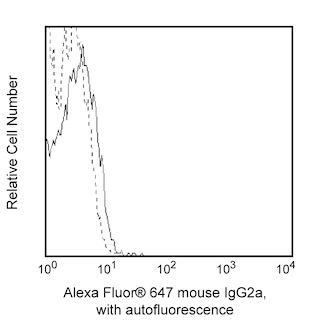
- An isotype control should be used at the same concentration as the antibody of interest.
- Caution: Sodium azide yields highly toxic hydrazoic acid under acidic conditions. Dilute azide compounds in running water before discarding to avoid accumulation of potentially explosive deposits in plumbing.
- Source of all serum proteins is from USDA inspected abattoirs located in the United States.
- The Alexa Fluor®, Pacific Blue™, and Cascade Blue® dye antibody conjugates in this product are sold under license from Molecular Probes, Inc. for research use only, excluding use in combination with microarrays, or as analyte specific reagents. The Alexa Fluor® dyes (except for Alexa Fluor® 430), Pacific Blue™ dye, and Cascade Blue® dye are covered by pending and issued patents.
- Alexa Fluor® is a registered trademark of Molecular Probes, Inc., Eugene, OR.
- Alexa Fluor® 647 fluorochrome emission is collected at the same instrument settings as for allophycocyanin (APC).
- For fluorochrome spectra and suitable instrument settings, please refer to our Multicolor Flow Cytometry web page at www.bdbiosciences.com/colors.
- Please refer to www.bdbiosciences.com/us/s/resources for technical protocols.
Companion Products


The CAM5.2 monoclonal antibody specifically recognizes cytokeratin having a primary reactivity with human keratin proteins that correspond to Moll's peptides #7 (48 kDa) and #8 (52 kDa). Cytokeratin 7 and 8 are type II cytoskeletal keratins. These cytoskeletal proteins provide structural integrity for epithelial cells and may serve other functions as well. They are expressed in epithelia cells that comprise normal human tissues. Although these cytokeratins are not normally expressed in stratified squamous epithelium, they may be expressed in some squamous cell carcinomas. The CAM 5.2 antibody stains most epithelial-derived tissue, including liver, renal tubular epithelium, and hepatocellular and renal cell carcinomas.
Development References (6)
-
Battifora H. Diagnostic uses of antibodies to keratins: A review and immunohistochemical comparison of seven monoclonal and three polyclonal antibodies. In: Fenoglio-Preiser C, Wolff M, Rilke F, ed. Progress in Surgical Pathology. 1988:1-15.
-
Cooper D, Schermer A, Sun TT. Biology of disease classification of human epithelia and their neoplasms using monoclonal antibodies to keratins: strategies, applications and limitations. Lab Invest. 1985; 52:243-256. (Clone-specific). View Reference
-
Leader M, Patel J, Makin C, Henry K. An analysis of the sensitivity and specificity of the cytokeratin marker CAM 5.2 for epithelial tumors: results of a study of 203 sarcomas, 50 carcinomas, and 28 malignant melanomas. Histopathology. 1986; 10:1315-1324. (Clone-specific: Immunohistochemistry). View Reference
-
Makin C, Bobrow L, Bodmer W. Monoclonal antibody to cytokeratin for use in routine histopathology. J Clin Pathol. 1984; 37(9):975-983. (Immunogen: Immunofluorescence, Immunohistochemistry, Immunoprecipitation, Western blot). View Reference
-
Moll R, Franke W, Schiller D, Geiger B, Krepler R. The catalog of human cytokeratins: patterns of expression in normal epithelia, tumors, and cultured cells. Cell. 1982; 31:11-24. (Clone-specific). View Reference
-
Smedts F, Ramaekers F, Robben H, et al. Changing patterns of keratin expression during progression of cervical intraepithelial neoplasia. Am J Pathol. 1990; 136(3):657-668. (Clone-specific: Immunohistochemistry, Western blot). View Reference
Please refer to Support Documents for Quality Certificates
Global - Refer to manufacturer's instructions for use and related User Manuals and Technical data sheets before using this products as described
Comparisons, where applicable, are made against older BD Technology, manual methods or are general performance claims. Comparisons are not made against non-BD technologies, unless otherwise noted.
For Research Use Only. Not for use in diagnostic or therapeutic procedures.