-
Reagents
- Flow Cytometry Reagents
-
Western Blotting and Molecular Reagents
- Immunoassay Reagents
-
Single-Cell Multiomics Reagents
- BD® OMICS-Guard Sample Preservation Buffer
- BD® AbSeq Assay
- BD® Single-Cell Multiplexing Kit
- BD Rhapsody™ ATAC-Seq Assays
- BD Rhapsody™ Whole Transcriptome Analysis (WTA) Amplification Kit
- BD Rhapsody™ TCR/BCR Next Multiomic Assays
- BD Rhapsody™ Targeted mRNA Kits
- BD Rhapsody™ Accessory Kits
- BD® OMICS-One Protein Panels
-
Functional Assays
-
Microscopy and Imaging Reagents
-
Cell Preparation and Separation Reagents
-
- BD® OMICS-Guard Sample Preservation Buffer
- BD® AbSeq Assay
- BD® Single-Cell Multiplexing Kit
- BD Rhapsody™ ATAC-Seq Assays
- BD Rhapsody™ Whole Transcriptome Analysis (WTA) Amplification Kit
- BD Rhapsody™ TCR/BCR Next Multiomic Assays
- BD Rhapsody™ Targeted mRNA Kits
- BD Rhapsody™ Accessory Kits
- BD® OMICS-One Protein Panels
- Poland (English)
-
Change country/language
Old Browser
This page has been recently translated and is available in French now.
Looks like you're visiting us from United States.
Would you like to stay on the current country site or be switched to your country?
BD Transduction Laboratories™ Purified Mouse Anti-CD49c
Clone 42/CD49c (RUO)


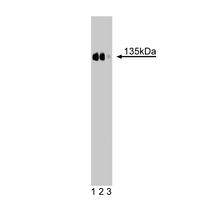
Western blot analysis of CD49c (integrin α3) on a rat kidney lysate. Lane 1: 1:250, lane 2: 1:500, lane 3: 1:1000 dilution of the mouse anti-CD49c antibody.

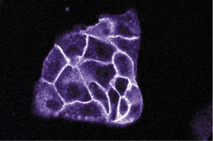
Immunofluorescence staining of MDCK cells (canine kidney; ATCC CCL-34).


BD Transduction Laboratories™ Purified Mouse Anti-CD49c

BD Transduction Laboratories™ Purified Mouse Anti-CD49c

Regulatory Status Legend
Any use of products other than the permitted use without the express written authorization of Becton, Dickinson and Company is strictly prohibited.
Preparation And Storage
Recommended Assay Procedures
Western blot: Please refer to http://www.bdbiosciences.com/pharmingen/protocols/Western_Blotting.shtml
Product Notices
- Since applications vary, each investigator should titrate the reagent to obtain optimal results.
- Please refer to www.bdbiosciences.com/us/s/resources for technical protocols.
- Caution: Sodium azide yields highly toxic hydrazoic acid under acidic conditions. Dilute azide compounds in running water before discarding to avoid accumulation of potentially explosive deposits in plumbing.
- Source of all serum proteins is from USDA inspected abattoirs located in the United States.
Data Sheets
Companion Products
.png?imwidth=320)

Integrins are a family of dimeric proteins that mediate cell-to-cell and extracellular matrix adhesion. They consist of a large α chain that is non-covalently associated with a smaller β chain which defines the integrin subfamilies. VLA-3 (Very Late Antigen-3), a member of the integrin superfamily, exhibits elevated expression on B lymphocytes, but is also found on monocytes, platelets, and hematopoietic progenitor cells. A heterodimer of α3 (CD49c) and β1 (CD29) subunits, VLA is a receptor for laminin, fibronectin, and collagen. The α3 chain contains a large extracellular domain with three putative metal-binding sequences, a transmembrane domain, and a short cytoplasmic tail. Differing requirements for divalent cations and the influence of RGD peptides results in multiple ligand-binding mechanisms for VLA-3. Although its expression is restricted in normal tissues, VLA-3 is found on a variety of cultured tumor cells. In addition, levels of VLA-3 have been shown to correlate with the degree of invasiveness of malignant melanoma cells. Thus, VLA-3 mediates intercellular adhesion and cell migration in normal and, possibly, cancerous cell types.
Development References (2)
-
Takada Y, Murphy E, Pil P, Chen C, Ginsberg MH, Hemler ME. Molecular cloning and expression of the cDNA for alpha 3 subunit of human alpha 3 beta 1 (VLA-3), an integrin receptor for fibronectin, laminin, and collagen. J Cell Physiol. 1991; 115(1):257-266. (Biology). View Reference
-
Takeuchi K, Hirano K, Tsuji T, Osawa T, Irimura T. cDNA cloning of mouse VLA-3 alpha subunit. J Cell Biochem. 1995; 57(2):371-377. (Biology). View Reference
Please refer to Support Documents for Quality Certificates
Global - Refer to manufacturer's instructions for use and related User Manuals and Technical data sheets before using this products as described
Comparisons, where applicable, are made against older BD Technology, manual methods or are general performance claims. Comparisons are not made against non-BD technologies, unless otherwise noted.
For Research Use Only. Not for use in diagnostic or therapeutic procedures.