


BD Horizon™ RB780 Mouse Anti-Human CD62L (L-Selectin)
克隆 DREG-56 (RUO)


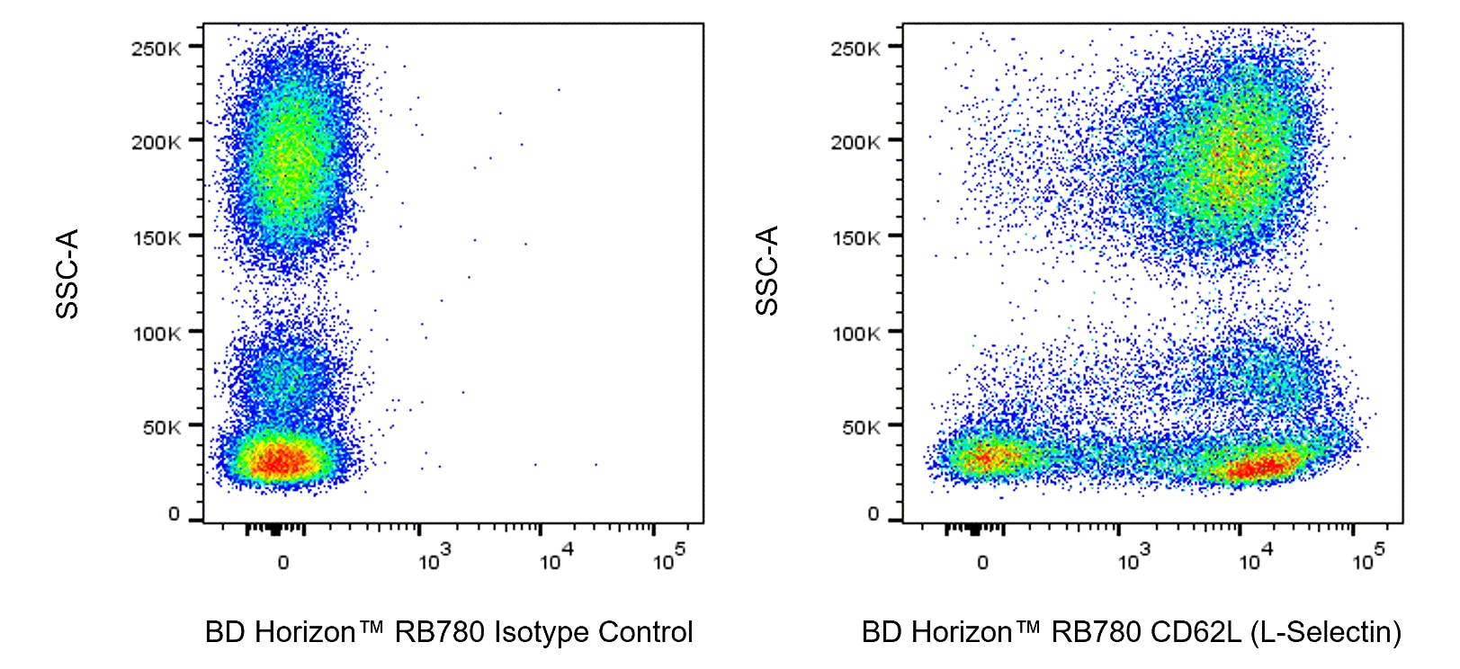
Multiparameter flow cytometric analysis of CD62L (L-Selectin) expression on Human peripheral blood leucocyte populations. Human whole blood was first treated with BD Pharm Lyse™ Lysing Buffer (Cat. No. 555899) to lyse erythrocytes, washed, and then preincubated with BD Pharmingen™ Human BD Fc Block™ (Cat. No. 564219/564220). The leucocytes were then stained with either BD Horizon™ RB780 Mouse IgG1, κ Isotype Control (Cat. No. 568532; Left Plot) or BD Horizon™ RB780 Mouse Anti-Human CD62L (L-Selectin) antibody (Cat. No. 569211/569212; Right Plot). The bivariate pseudocolor density plot showing the correlated expression of CD62L (L-Selectin) [or Ig Isotype control staining] versus side light-scatter (SSC-A) signals was derived from gated events with the forward and side light-scatter characteristics of viable leucocytes. Flow cytometry and data analysis were performed using a BD LSRFortessa™ X-20 Cell Analyzer System and FlowJo™ software.

Multiparameter flow cytometric analysis of CD62L (L-Selectin) expression on Human peripheral blood leucocyte populations. Human whole blood was first treated with BD Pharm Lyse™ Lysing Buffer (Cat. No. 555899) to lyse erythrocytes, washed, and then preincubated with BD Pharmingen™ Human BD Fc Block™ (Cat. No. 564219/564220). The leucocytes were then stained with either BD Horizon™ RB780 Mouse IgG1, κ Isotype Control (Cat. No. 568532; Left Plot) or BD Horizon™ RB780 Mouse Anti-Human CD62L (L-Selectin) antibody (Cat. No. 569211/569212; Right Plot). The bivariate pseudocolor density plot showing the correlated expression of CD62L (L-Selectin) [or Ig Isotype control staining] versus side light-scatter (SSC-A) signals was derived from gated events with the forward and side light-scatter characteristics of viable leucocytes. Flow cytometry and data analysis were performed using a BD LSRFortessa™ X-20 Cell Analyzer System and FlowJo™ software.


准备和存储


推荐的实验流程
BD® CompBeads can be used as surrogates to assess fluorescence spillover (compensation). When fluorochrome conjugated antibodies are bound to BD® CompBeads, they have spectral properties very similar to cells. However, for some fluorochromes there can be small differences in spectral emissions compared to cells, resulting in spillover values that differ when compared to biological controls. It is strongly recommended that when using a reagent for the first time, users compare the spillover on cells and BD® CompBeads to ensure that BD® CompBeads are appropriate for your specific cellular application.
商品通知
- When using high concentrations of antibody, background binding of this dye to erythroid fragments produced by ammonium chloride-based lysis, such as with BD Pharm Lyse™ Lysing Buffer (Cat. No. 555899), has been observed when the antibody conjugate was present during the lysis procedure. This may cause nonspecific staining of target cells, such as leukocytes, which have bound the resulting erythroid fragments. This background can be mitigated by any of the following: titrating the antibody conjugate to a lower concentration, fixing samples with formaldehyde, or removing erythrocytes before staining (eg, gradient centrifugation or pre-lysis with wash). This background has not been observed when cells were lysed with BD FACS™ Lysing Solution (Cat. No. 349202) after staining.
- Please refer to www.bdbiosciences.com/us/s/resources for technical protocols.
- This reagent has been pre-diluted for use at the recommended Volume per Test. We typically use 1 × 10^6 cells in a 100-µl experimental sample (a test).
- An isotype control should be used at the same concentration as the antibody of interest.
- Please observe the following precautions: Absorption of visible light can significantly alter the energy transfer occurring in any tandem fluorochrome conjugate; therefore, we recommend that special precautions be taken (such as wrapping vials, tubes, or racks in aluminum foil) to prevent exposure of conjugated reagents, including cells stained with those reagents, to room illumination.
- Caution: Sodium azide yields highly toxic hydrazoic acid under acidic conditions. Dilute azide compounds in running water before discarding to avoid accumulation of potentially explosive deposits in plumbing.
- For fluorochrome spectra and suitable instrument settings, please refer to our Multicolor Flow Cytometry web page at www.bdbiosciences.com/colors.
- Human donor specific background has been observed in relation to the presence of anti-polyethylene glycol (PEG) antibodies, developed as a result of certain vaccines containing PEG, including some COVID-19 vaccines. We recommend use of BD Horizon Brilliant™ Stain Buffer in your experiments to help mitigate potential background. For more information visit https://www.bdbiosciences.com/en-us/support/product-notices.
- Please refer to http://regdocs.bd.com to access safety data sheets (SDS).
- Cy is a trademark of Global Life Sciences Solutions Germany GmbH or an affiliate doing business as Cytiva.
- For U.S. patents that may apply, see bd.com/patents.
配套商品



The DREG-56 monoclonal antibody specifically binds to CD62L. CD62L is a 76-95 kDa glycoprotein that is also referred to as L-selectin or LECAM-1. CD62L is expressed on neutrophils, monocytes, T- and B-lymphocyte subsets and NK cells. The DREG-56 antibody recognizes the same antigen as LAM-1, and specifically inhibits >90% of binding of human lymphocytes to high endothelial venules (HEV) in frozen sections of peripheral, but not mucosal lymphoid tissue. It thus defines L-selectin as a human lymphocyte homing receptor for peripheral lymph node HEV.

研发参考 (5)
-
Ivetic A, Hoskins Green HL, Hart SJ. L-selectin: A Major Regulator of Leukocyte Adhesion, Migration and Signaling.. Front Immunol. 2019; 10:1068. (Biology). 查看参考
-
Kishimoto TK, Jutila MA, Butcher EC. Identification of a human peripheral lymph node homing receptor: a rapidly down-regulated adhesion molecule. Proc Natl Acad Sci U S A. 1990; 87(6):2244-2248. (Clone-specific: Inhibition). 查看参考
-
Kishimoto TK, Warnock RA, Jutila MA, et al. Antibodies against human neutrophil LECAM-1 (LAM-1/Leu-8/DREG-56 antigen) and endothelial cell ELAM-1 inhibit a common CD18-independent adhesion pathway in vitro. Blood. 1991; 78(3):805-811. (Immunogen: Flow cytometry, Inhibition). 查看参考
-
Polley MJ, Phillips ML, Wayner E, et al. CD62 and endothelial cell-leukocyte adhesion molecule 1 (ELAM-1) recognize the same carbohydrate ligand, sialyl-Lewis x. Proc Natl Acad Sci U S A. 1991; 88(14):6224-6228. (Biology). 查看参考
-
Schlossman SF. Stuart F. Schlossman .. et al., ed. Leucocyte typing V : white cell differentiation antigens : proceedings of the fifth international workshop and conference held in Boston, USA, 3-7 November, 1993. Oxford: Oxford University Press; 1995.
Please refer to Support Documents for Quality Certificates
Global - Refer to manufacturer's instructions for use and related User Manuals and Technical data sheets before using this products as described
Comparisons, where applicable, are made against older BD Technology, manual methods or are general performance claims. Comparisons are not made against non-BD technologies, unless otherwise noted.
For Research Use Only. Not for use in diagnostic or therapeutic procedures.