Old Browser
Looks like you're visiting us from {countryName}.
Would you like to stay on the current country site or be switched to your country?


Analysis of LAT (pY226) in activated human T leukemia cells. Jurkat cells (ATCC TIB152) were serum starved overnight and then either stimulated with 5 mM hydrogen peroxide for 15 minutes (shaded histogram) or unstimulated (open histogram). The cells were fixed using BD Phosflow™ Fix Buffer I (Cat. No. 557870) for 10 minutes at 37°C, then permeabilized using BD Phosflow™ Perm Buffer III (Cat. No. 558050) on ice for at least 30 minutes, and then stained with Alexa Fluor® 647 Mouse anti-LAT (pY226) (Cat. No. 558432). Flow cytometry was performed on a BD FACSCalibur™ flow cytometry system.


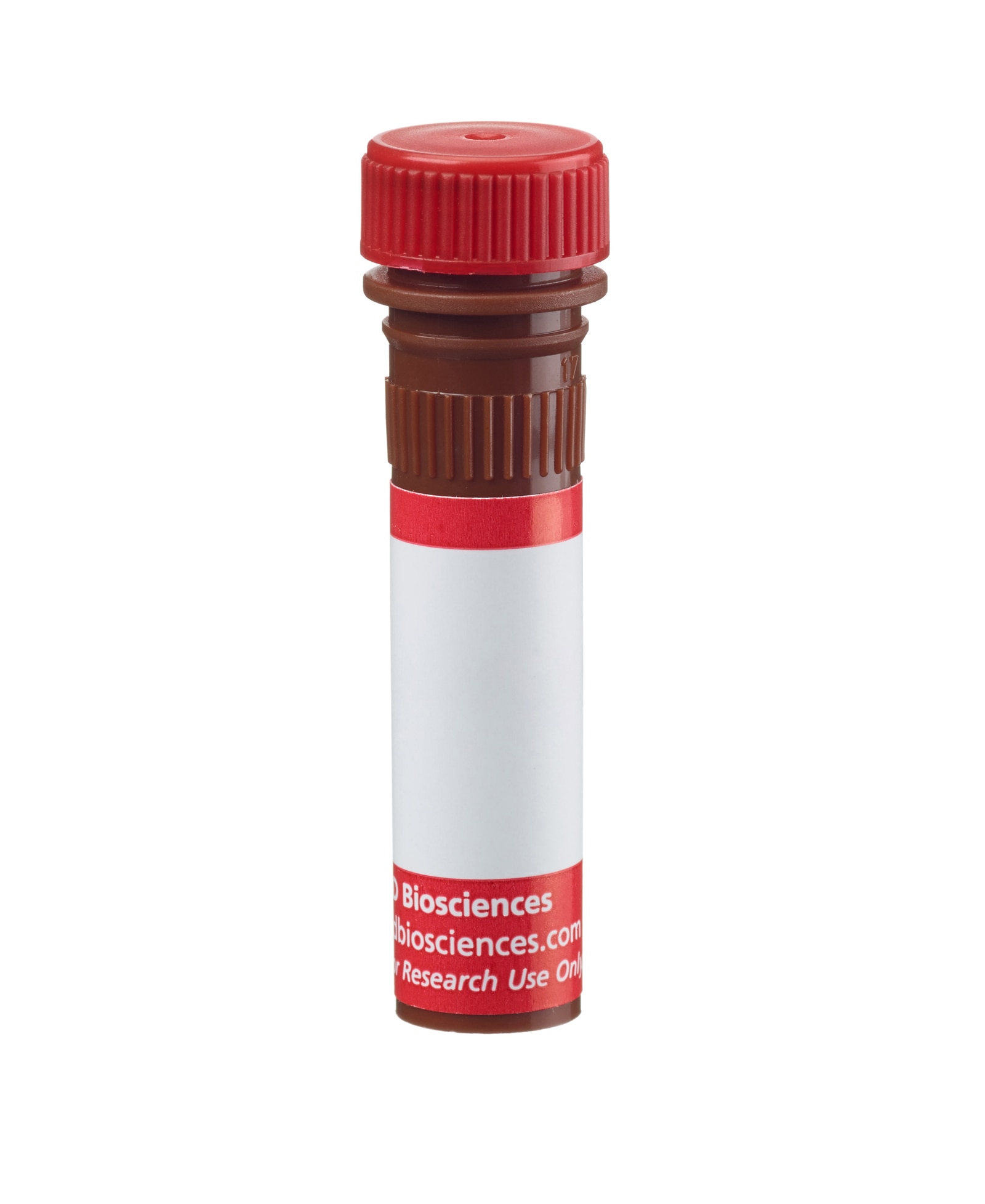
BD™ Phosflow Alexa Fluor® 647 Mouse anti-LAT (pY226)

监管状态图例
未经BD明确书面授权,严禁使用未经许可的任何商品。
准备和存储
商品通知
- This reagent has been pre-diluted for use at the recommended Volume per Test. We typically use 1 × 10^6 cells in a 100-µl experimental sample (a test).
- Please refer to www.bdbiosciences.com/us/s/resources for technical protocols.
- The Alexa Fluor®, Pacific Blue™, and Cascade Blue® dye antibody conjugates in this product are sold under license from Molecular Probes, Inc. for research use only, excluding use in combination with microarrays, or as analyte specific reagents. The Alexa Fluor® dyes (except for Alexa Fluor® 430), Pacific Blue™ dye, and Cascade Blue® dye are covered by pending and issued patents.
- Alexa Fluor® 647 fluorochrome emission is collected at the same instrument settings as for allophycocyanin (APC).
- Alexa Fluor® is a registered trademark of Molecular Probes, Inc., Eugene, OR.
- Caution: Sodium azide yields highly toxic hydrazoic acid under acidic conditions. Dilute azide compounds in running water before discarding to avoid accumulation of potentially explosive deposits in plumbing.
- Source of all serum proteins is from USDA inspected abattoirs located in the United States.
- For fluorochrome spectra and suitable instrument settings, please refer to our Multicolor Flow Cytometry web page at www.bdbiosciences.com/colors.
Engagement of the T cell receptor (TCR) induces signal transduction pathways that enhance gene transcription and cellular proliferation and differentiation. TCR ligation results in the recruitment and activation of multiple protein tyrosine kinases (PTKs), including lck, fyn, and ZAP70. Adaptor proteins, such as Grb2 and SLP-76, relay the signal to downstream effector molecules. LAT (linker for activation of T cells) is a substrate of the activated ZAP70 and functions to bridge the activated TCR and its associated PTKs with tyrosine kinase substrates. LAT is expressed as 36- and 38-kDa forms that result from post-translational modification, and as a 42-kDa form that results from alternative splicing. LAT is an integral membrane protein that is phosphorylated at five tyrosine sites upon TCR ligation. Following phosphorylation, LAT binds a number of important signaling molecules, including Grb2, Vav, PLCγ1, and the p85 subunit of PI3K. Multiple studies have shown that functional LAT is required for T lymphocyte activation and thymocyte development.
The J96-1238.58.93 monoclonal antibody recognizes the phosphorylated tyrosine 226 (pY226) of LAT, which is one of the phosphotyrosine sites required for binding Vav, Grb2, and Gads.
研发参考 (5)
-
Janssen E, Zhang W. Adaptor proteins in lymphocyte activation. Curr Opin Immunol. 2003; 15:269-276. (Biology). 查看参考
-
Lin J, Weiss A. Identification of the minimal tyrosine residues required for linker for activation of T cell function. J Biol Chem. 2001; 276:29588-29595. (Biology). 查看参考
-
Paz PE, Wang S, Clarke H, Lu X, Stokoe D, Abo A. Mapping the ZAP-70 phosphorylation sites on LAT (linker for activation of T cells) required for recruitment and activation of signalling proteins in T cells. Biochem J. 2001; 356:461-471. (Biology). 查看参考
-
Samelson LE. Signal transduction mediated by the T cell antigen receptor: The role of adapter proteins. Annu Rev Immunol. 2002; 20:371-394. (Biology).
-
Zhu M, Janssen E, Zhang W. Minimal requirement of tyrosine residues of linker for activation of T cells in TCR signaling and thymocyte development. J Immunol. 2003; 170:325-333. (Biology). 查看参考
Please refer to Support Documents for Quality Certificates
Global - Refer to manufacturer's instructions for use and related User Manuals and Technical data sheets before using this products as described
Comparisons, where applicable, are made against older BD Technology, manual methods or are general performance claims. Comparisons are not made against non-BD technologies, unless otherwise noted.
For Research Use Only. Not for use in diagnostic or therapeutic procedures.
Report a Site Issue
This form is intended to help us improve our website experience. For other support, please visit our Contact Us page.