

Preparation And Storage
Product Notices
- Since applications vary, each investigator should titrate the reagent to obtain optimal results.
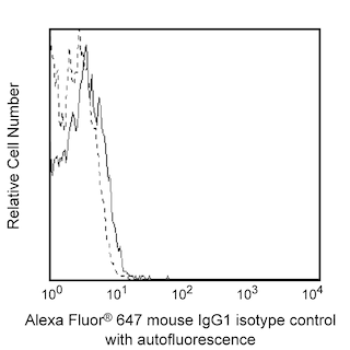
- An isotype control should be used at the same concentration as the antibody of interest.
- Caution: Sodium azide yields highly toxic hydrazoic acid under acidic conditions. Dilute azide compounds in running water before discarding to avoid accumulation of potentially explosive deposits in plumbing.
- Alexa Fluor® is a registered trademark of Molecular Probes, Inc., Eugene, OR.
- Alexa Fluor® 647 fluorochrome emission is collected at the same instrument settings as for allophycocyanin (APC).
- The Alexa Fluor®, Pacific Blue™, and Cascade Blue® dye antibody conjugates in this product are sold under license from Molecular Probes, Inc. for research use only, excluding use in combination with microarrays, or as analyte specific reagents. The Alexa Fluor® dyes (except for Alexa Fluor® 430), Pacific Blue™ dye, and Cascade Blue® dye are covered by pending and issued patents.
- For fluorochrome spectra and suitable instrument settings, please refer to our Multicolor Flow Cytometry web page at www.bdbiosciences.com/colors.
- Please refer to www.bdbiosciences.com/us/s/resources for technical protocols.
Companion Products


The OX-6 antibody specifically recognizes non-polymorphic determinants of the Rat MHC class II antigen, I-A equivalent. RT1B is found on peripheral B lymphocytes, thymic cortical epithelial and medullary reticular cells, small intestinal villus epithelium, epidermal Langerhans cells, dendritic cells, some tissue macrophage populations, peritoneal mast cells, and a subset of thymocytes, but not on peripheral T cells, erythrocytes, or microglia. The OX-6 mAb cross-reacts with mouse I-A[k] and I-A[s] alloantigens and with a major subset of splenocytes from NOD (I-A[g7]) mice.
Development References (10)
-
Chen-Woan M, Delaney CP, Fournier V, et al. In vitro characterization of rat bone marrow-derived dendritic cells and their precursors. J Leukoc Biol. 1996; 59(2):196-207. (Biology). View Reference
-
Damoiseaux JG, Yagita H, Okumura K, van Breda Vriesman PJ. Costimulatory molecules CD80 and CD86 in the rat; tissue distribution and expression by antigen-presenting cells. J Leukoc Biol. 1998; 64(6):803-809. (Biology). View Reference
-
Dick AD, Ford AL, Forrester JV, Sedgwick JD. Flow cytometric identification of a minority population of MHC class II positive cells in the normal rat retina distinct from CD45lowCD11b/c+CD4low parenchymal microglia. J Leukoc Biol. 1995; 79(9):834-840. (Biology). View Reference
-
Fox CC, Jewell SD, Whitacre CC. Rat peritoneal mast cells present antigen to a PPD-specific T cell line. Cell Immunol. 1994; 158(1):253-264. (Biology). View Reference
-
Fukumoto T, McMaster WR, Williams AF, et al. Mouse monoclonal antibodies against rat major histocompatibility antigens. Two Ia antigens and expression of Ia and class I antigens in rat thymus. Eur J Immunol. 1982; 12:237-243. (Biology). View Reference
-
Mayrhofer G, Pugh CW, Barclay AN. The distribution, ontogeny and origin in the rat of Ia-positive cells with dendritic morphology and of Ia antigen in epithelia, with special reference to the intestine. Eur J Immunol. 1983; 13(2):112-122. (Biology). View Reference
-
McMaster WR, Williams AF et al. Identification of Ia glycoproteins in rat thymus and purification from rat spleen.. Eur J Immunol. 1979; 9:426-433. (Immunogen). View Reference
-
Neiss U, Becker D, Knop J, Reske K. Modulation of MHC class II determinants on rat Langerhans cells during short term culture. Adv Exp Med Biol. 1993; 329:29-34. (Clone-specific). View Reference
-
Nelson DJ, McMenamin C, McWilliam AS, Brenan M, Holt PG. Development of the airway intraepithelial dendritic cell network in the rat from class II major histocompatibility (Ia)-negative precursors: differential regulation of Ia expression at different levels of the respiratory tract. J Exp Med. 1994; 179(1):203-212. (Biology). View Reference
-
Xia WJ, Schneeberger EE, McCarthy K, Kradin RL. Accessory cells of the lung. II. Ia+ pulmonary dendritic cells display cell surface antigen heterogeneity. Am J Respir Cell Mol Biol. 1991; 5(3):276-283. (Biology). View Reference
Please refer to Support Documents for Quality Certificates
Global - Refer to manufacturer's instructions for use and related User Manuals and Technical data sheets before using this products as described
Comparisons, where applicable, are made against older BD Technology, manual methods or are general performance claims. Comparisons are not made against non-BD technologies, unless otherwise noted.
For Research Use Only. Not for use in diagnostic or therapeutic procedures.