BUV805 Rat Anti-Mouse CD8a
Clone 53-6.7 (RUO)
-
Product Announcement
This product is the replacement for 564920
- Brand BD Horizon™
- Alternative Name Cd8a; CD8 alpha chain; Ly-2; Lyt2; Lyt-2; Ly-35; Ly-B
- Concentration 0.2 mg/ml
- Isotype Rat LOU, also known as Louvain, LOU/C, LOU/M IgG2a, κ
- Reactivity Mouse (QC Testing)
- Application
Flow cytometry (Routinely Tested)
- Immunogen Mouse Spleen Cells or Thymocyte Membranes
- Entrez Gene ID 12525
- Storage Buffer Aqueous buffered solution containing ≤0.09% sodium azide.
- Regulatory Status RUO
Regulatory Status Legend
Description
The 53-6.7 monoclonal antibody specifically binds to the 38 kDa α and 34 kDa α' chains of the CD8 differentiation antigen (Ly-2 or Lyt-2) of all mouse strains tested. The CD8 α and α' chains (CD8a) form heterodimers with the CD8 β chain (CD8b, Ly-3, or Lyt-3) on the surface of most thymocytes. A subpopulation of mature T lymphocytes (i.e., MHC class I-restricted T cells, including most T suppressor/cytotoxic cells) expresses almost exclusively the CD8 αβ heterodimer. Subsets of γδ TCR-bearing T cells, intestinal intrapithelial lymphocytes, and dendritic cells express CD8a without CD8b. It has been suggested that the expression of the CD8a/CD8b heterodimer is restricted to T lymphocytes which matured in the thymus or in an extrathymic environment that had been influenced by thymus-initiated neuroendocrine signals. CD8 is an antigen coreceptor on the T-cell surface which interacts with MHC class I molecules on antigen-presenting cells or epithelial cells. It participates in T-cell activation through its association with the T-cell receptor complex and protein tyrosine kinase lck (p56 [lck]). The CD8 α and α' chains arise from alternatively spliced messengers of a single CD8a gene. The longer α form associates with p56 [lck] via a CXCP motif in its cytoplasmic domain, which it shares with CD4, but not with CD8b. The truncated α' chain is unable to associate with p56 [lck], and it may function to attenuate the CD8-mediated costimulatory signal during intrathymic T-cell maturation. In vivo and in vitro treatment with 53-6.7 mAb has reportedly been effective at depleting CD8+ peripheral T lymphocytes. The 53-6.7 antibody has also been reported to cross-react with CD8 α- and α'-like polypeptides on subsets of thymic and peripheral lymphocytes in the Egyptian toad, Bufo regularis.
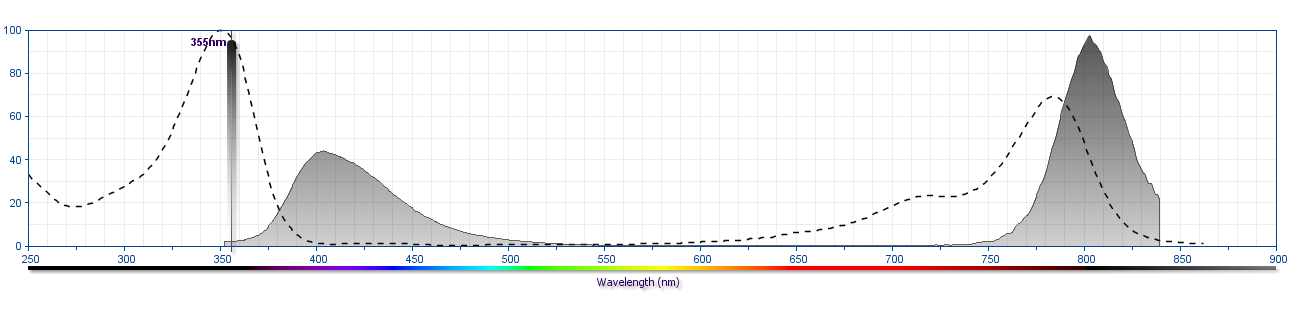
The antibody was conjugated to BD Horizon BUV805 which is part of the BD Horizon Brilliant™ Ultraviolet family of dyes. This dye is a tandem fluorochrome with an Ex Max near 350 nm and an Em Max near 805 nm. BD Horizon Brilliant BUV805 can be excited by the ultraviolet laser (355 nm) and detected with a 820/60 nm filter and a 770 nm LP.
Format
BUV805 is a tandem fluorochrome that combines BD Horizon BUV395 and an acceptor dye with an Em Max at 805 nm. BUV805 has been exclusively developed by BD Biosciences for instruments equipped with a 355-nm UV laser.
Other Formats →
Suggested Companion Products
Brilliant Stain Buffer RUO
100 Tests
Cat No: 563794
Brilliant Stain Buffer RUO
1000 Tests
Cat No: 566349
Brilliant Stain Buffer Plus RUO
1000 Tests
Cat No: 566385
Purified Rat Anti-Mouse CD16/CD32 (Mouse BD Fc Block™) 2.4G2 RUO
0.1 mg
Cat No: 553141
Purified Rat Anti-Mouse CD16/CD32 (Mouse BD Fc Block™) 2.4G2 RUO
0.5 mg
Cat No: 553142
Lysing Buffer RUO
100 mL
Cat No: 555899
BUV805 Rat IgG2a, κ Isotype Control R35-95 RUO
50 µg
Cat No: 612899
Stain Buffer (FBS) RUO
500 mL
Cat No: 554656
Stain Buffer (BSA) RUO
500 mL
Cat No: 554657
Resources & Tools | ||||||
---|---|---|---|---|---|---|
Spectrum Viewer | Panel Designer | Spectrum Viewer | Download TDS | Regulatory Document Website |
Preparation and Storage
Store undiluted at 4°C and protected from prolonged exposure to light. Do not freeze.
The monoclonal antibody was purified from tissue culture supernatant or ascites by affinity chromatography.
The antibody was conjugated with BD Horizon BUV805 under optimum conditions, and unconjugated antibody and free BD Horizon BUV805 were removed.
Product Notices
- Since applications vary, each investigator should titrate the reagent to obtain optimal results.
- An isotype control should be used at the same concentration as the antibody of interest.
- Caution: Sodium azide yields highly toxic hydrazoic acid under acidic conditions. Dilute azide compounds in running water before discarding to avoid accumulation of potentially explosive deposits in plumbing.
- For fluorochrome spectra and suitable instrument settings, please refer to our Multicolor Flow Cytometry web page at www.bdbiosciences.com/colors.
- BD Horizon Brilliant Ultraviolet 805 is covered by one or more of the following US patents: 8,110,673, 8,158,444; 8,227,187; 8,575,303; 8,354,239.
- BD Horizon Brilliant Stain Buffer is covered by one or more of the following US patents: 8,110,673; 8,158,444; 8,575,303; 8,354,239.
- Please refer to www.bdbiosciences.com/us/s/resources for technical protocols.
BD™ CompBeads can be used as surrogates to assess fluorescence spillover (Compensation). When fluorochrome-conjugated antibodies are bound to BD CompBeads, they have spectral properties very similar to cells. However, for some fluorochromes there can be small differences in spectral emissions compared to cells, resulting in spillover values that differ when compared to biological controls. It is strongly recommended that when using a reagent for the first time, users compare the spillover on cells and BD CompBeads. This will ensure that BD CompBeads are appropriate for your specific cellular application.
For optimal and reproducible results, BD Horizon Brilliant Stain Buffer should be used anytime two or more BD Horizon Brilliant dyes are used in the same experiment. Fluorescent dye interactions may cause staining artifacts which may affect data interpretation. The BD Horizon Brilliant Stain Buffer was designed to minimize these interactions. More information can be found in the Technical Data Sheet of the BD Horizon Brilliant Stain Buffer (Cat. No. 563794/566349) or the BD Horizon Brilliant Stain Buffer Plus (Cat. No. 566385).
Note: When using high concentrations of antibody, background binding of this dye to erythroid cell subsets (mature erythrocytes and precursors) has been observed. For researchers studying these cell populations, or in cases where light scatter gating does not adequately exclude these cells from the analysis, this background may be an important factor to consider when selecting reagents for panel(s).