Preparation And Storage
Product Notices
- Since applications vary, each investigator should titrate the reagent to obtain optimal results.
- An isotype control should be used at the same concentration as the antibody of interest.
- Alexa Fluor® is a registered trademark of Molecular Probes, Inc., Eugene, OR.
- Caution: Sodium azide yields highly toxic hydrazoic acid under acidic conditions. Dilute azide compounds in running water before discarding to avoid accumulation of potentially explosive deposits in plumbing.
- For fluorochrome spectra and suitable instrument settings, please refer to our Multicolor Flow Cytometry web page at www.bdbiosciences.com/colors.
- Please refer to http://regdocs.bd.com to access safety data sheets (SDS).
- Please refer to www.bdbiosciences.com/us/s/resources for technical protocols.
Companion Products


.png?imwidth=320)

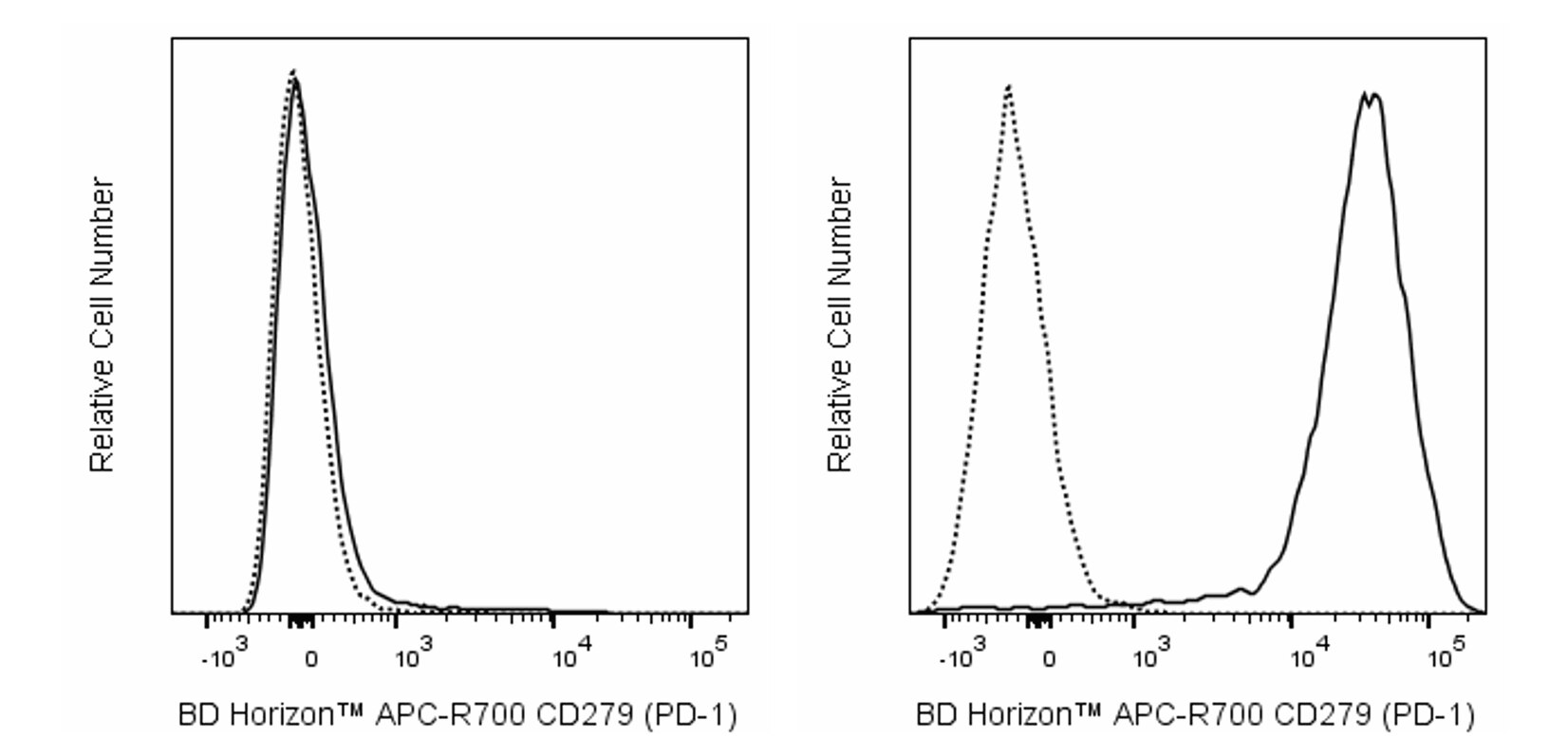
The RMP1-30 monoclonal antibody specifically recognizes CD279 which is also known as PD-1 (programmed death-1). CD279 (PD-1) is a ~55 kDa type I transmembrane glycoprotein that is encoded by Pdcd1 which belongs to the CD28/CTLA-4 family within the Ig superfamily. CD279 (PD-1) is comprised of an extracellular region with an IgV-like domain and an intracellular region with an immunoreceptor tyrosine-based inhibitory motif (ITIM) and an immunoreceptor tyrosine-based switch motif (ITSM) that are associated with inhibitory signaling functions. CD279 (PD-1) is transiently expressed on CD4-CD8- thymocytes and developing B lymphocytes at the pro-B-cell stage. It is also expressed on activated myeloid cells, B cells, and T cells including exhausted T cells found in mice during chronic viral infections or cancer. This co-inhibitory receptor reportedly functions in negative regulation of immune responses and thus helps guard against autoimmunity and preserves peripheral tolerance. CD273 (also known as PD-L2 or B7-DC) and CD274 (PD-L1 or B7-H1) are members of the B7 family within the Ig superfamily that serve as ligands for CD279 (PD-1). The RMP1-30 antibody reportedly does not block the binding of CD279 (PD-1) to these ligands.
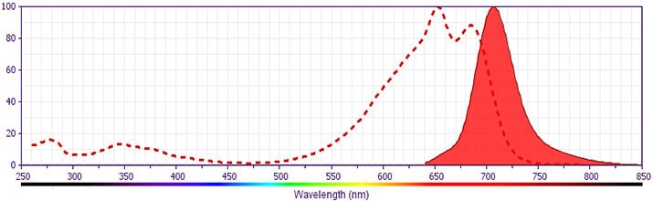
This antibody was conjugated to BD Horizon APC-R700, which has been developed exclusively by BD Biosciences as a better alternative to Alexa Fluor® 700. APC-R700 excites and emits at similar wavelengths to Alexa Fluor® 700 yet exhibits significantly improved brightness. This dye can be excited by the red laser and detected with the same filter set as Alexa Fluor® (eg, 730/45-nm filter).
Development References (7)
-
Agata Y, Kawasaki A, Nishimura H, et al. Expression of the PD-1 antigen on the surface of stimulated mouse T and B lymphocytes. Int Immunol. 1996 May; 8(5):765-772. (Biology). View Reference
-
Gupta PK, Godec J, Wolski D, et al. CD39 Expression Identifies Terminally Exhausted CD8+ T Cells. PLoS Pathogens. 2015; 11(10):e1005177. (Clone-specific: Flow cytometry). View Reference
-
Kasagi S, Kawano S, Okazaki T, et al. Anti-programmed cell death 1 antibody reduces CD4+PD-1+ T cells and relieves the lupus-like nephritis of NZB/W F1 mice. J Immunol. 2010; 184(5):2337-2347. (Clone-specific: Flow cytometry). View Reference
-
Liu X, Gibbons RM, Harrington SM, et al. Endogenous tumor-reactive CD8(+) T cells are differentiated effector cells expressing high levels of CD11a and PD-1 but are unable to control tumor growth. Oncoimmunology. 2013; 2(6):e23972. (Clone-specific: Flow cytometry). View Reference
-
Matsumoto K, Inoue H, Nakano T, et al. B7-DC regulates asthmatic response by an IFN-gamma-dependent mechanism. J Immunol. 2004; 172(4):2530-2541. (Immunogen: Flow cytometry, Functional assay). View Reference
-
Nishimura H, Agata Y, Kawasaki A, et al. Developmentally regulated expression of the PD-1 protein on the surface of double-negative (CD4-CD8-) thymocytes. Int Immunol. 1996 May; 8(5):773-780. (Biology). View Reference
-
Tsushima F, Iwai H, Otsuki N, et al. Preferential contribution of B7-H1 to programmed death-1-mediated regulation of hapten-specific allergic inflammatory responses. Eur J Immunol. 2003; 33(10):2773-2782. (Biology). View Reference
Please refer to Support Documents for Quality Certificates
Global - Refer to manufacturer's instructions for use and related User Manuals and Technical data sheets before using this products as described
Comparisons, where applicable, are made against older BD Technology, manual methods or are general performance claims. Comparisons are not made against non-BD technologies, unless otherwise noted.
For Research Use Only. Not for use in diagnostic or therapeutic procedures.