
Preparation And Storage
Recommended Assay Procedures
BD™ CompBeads can be used as surrogates to assess fluorescence spillover (Compensation). When fluorochrome conjugated antibodies are bound to CompBeads, they have spectral properties very similar to cells. However, for some fluorochromes there can be small differences in spectral emissions compared to cells, resulting in spillover values that differ when compared to biological controls. It is strongly recommended that when using a reagent for the first time, users compare the spillover on cell and CompBead to ensure that BD Comp beads are appropriate for your specific cellular application.
Product Notices
- Since applications vary, each investigator should titrate the reagent to obtain optimal results.
- An isotype control should be used at the same concentration as the antibody of interest.
- Caution: Sodium azide yields highly toxic hydrazoic acid under acidic conditions. Dilute azide compounds in running water before discarding to avoid accumulation of potentially explosive deposits in plumbing.
- Alexa Fluor® is a registered trademark of Molecular Probes, Inc., Eugene, OR.
- Alexa Fluor® 647 fluorochrome emission is collected at the same instrument settings as for allophycocyanin (APC).
- The Alexa Fluor®, Pacific Blue™, and Cascade Blue® dye antibody conjugates in this product are sold under license from Molecular Probes, Inc. for research use only, excluding use in combination with microarrays, or as analyte specific reagents. The Alexa Fluor® dyes (except for Alexa Fluor® 430), Pacific Blue™ dye, and Cascade Blue® dye are covered by pending and issued patents.
- For fluorochrome spectra and suitable instrument settings, please refer to our Multicolor Flow Cytometry web page at www.bdbiosciences.com/colors.
- This product is provided under an intellectual property license between Life Technologies Corporation and BD Businesses. The purchase of this product conveys to the buyer the non-transferable right to use the purchased amount of the product and components of the product in research conducted by the buyer (whether the buyer is an academic or for-profit entity). The buyer cannot sell or otherwise transfer (a) this product (b) its components or (c) materials made using this product or its components to a third party or otherwise use this product or its components or materials made using this product or its components for Commercial Purposes. Commercial Purposes means any activity by a party for consideration and may include, but is not limited to: (1) use of the product or its components in manufacturing; (2) use of the product or its components to provide a service, information, or data; (3) use of the product or its components for therapeutic, diagnostic or prophylactic purposes; or (4) resale of the product or its components, whether or not such product or its components are resold for use in research. For information on purchasing a license to this product for any other use, contact Life Technologies Corporation, Cell Analysis Business Unit Business Development, 29851 Willow Creek Road, Eugene, OR 97402, USA, Tel: (541) 465-8300. Fax: (541) 335-0504.
- Please refer to http://regdocs.bd.com to access safety data sheets (SDS).
- Please refer to www.bdbiosciences.com/us/s/resources for technical protocols.
Companion Products


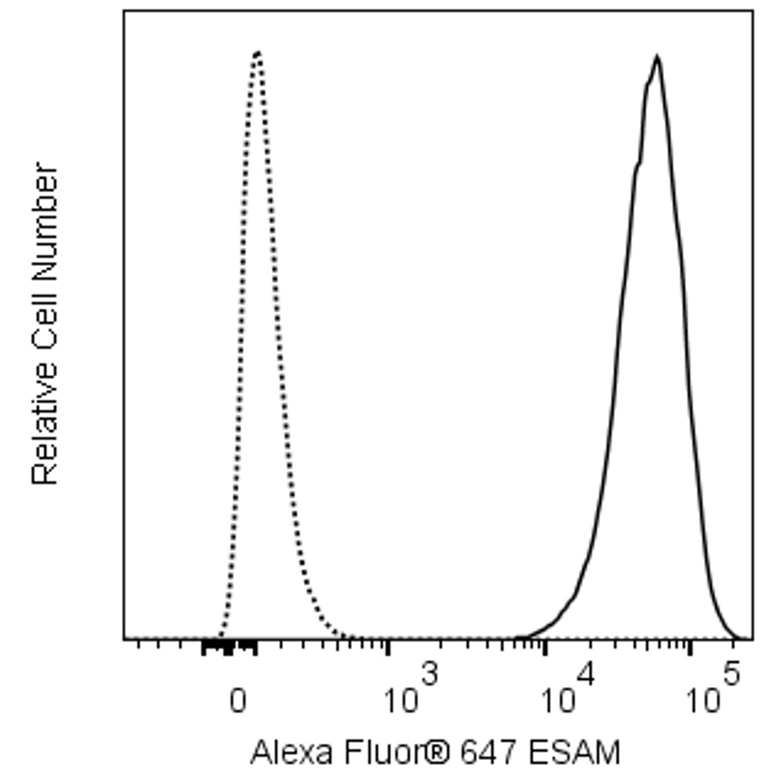
The 1G8 monoclonal antibody specifically recognizes Endothelial cell-selective adhesion molecule (ESAM). ESAM is a ~55 kDa single-pass type I transmembrane glycoprotein that is encoded by ESAM (Endothelial cell-specific adhesion molecule) which belongs to the immunoglobulin supergene superfamily (IgSF). ESAM contains an IgV-type and IgC2-type domain in its extracellular region followed by a transmembrane sequence and cytoplasmic tail. ESAM is strongly expressed on platelets, megakaryocytes, and endothelial cells at interendothelial cell junctions as well as dendritic cells. Through homophilic interactions, this interendothelial cell adhesion molecule may play roles in regulating vascular permeability and in the extravasation of leucocytes, eg, neutrophils, through blood vessel walls. ESAM is also reportedly expressed on hematopoietic stem cells (HSCs). ESAM-positive cell populations are enriched for multipotent myeloid erythroid progenitors and primitive progenitors with lymphopoietic activity.
Development References (7)
-
Duong CN, Nottebaum AF, Butz S, et al. Interference With ESAM (Endothelial Cell-Selective Adhesion Molecule) Plus Vascular Endothelial-Cadherin Causes Immediate Lethality and Lung-Specific Blood Coagulation. Arterioscler Thromb Vasc Biol. 2020; 40(2):378-393. (Clone-specific: Flow cytometry, Fluorescence microscopy, Immunofluorescence, Western blot). View Reference
-
Fujita K, Chakarov S, Kobayashi T, et al. Cell-autonomous FLT3L shedding via ADAM10 mediates conventional dendritic cell development in mouse spleen. Proc Natl Acad Sci U S A. 2019; 116(29):14714-14723. (Clone-specific: Flow cytometry). View Reference
-
Hirata Ki, Ishida T, Penta K, et al. Cloning of an immunoglobulin family adhesion molecule selectively expressed by endothelial cells.. J Biol Chem. 2001; 276(19):16223-31. (Biology). View Reference
-
Kirkling ME, Cytlak U, Lau CM, et al. Notch Signaling Facilitates In Vitro Generation of Cross-Presenting Classical Dendritic Cells. Cell Rep. 2018; 23(12):3658-3672. (Clone-specific: Flow cytometry). View Reference
-
Lewis KL, Caton ML, Bogunovic M, et al. Notch2 receptor signaling controls functional differentiation of dendritic cells in the spleen and intestine. Immunity. 2011; 35(5):780-791. (Biology). View Reference
-
Nasdala I, Wolburg-Buchholz K, Wolburg H, et al. A transmembrane tight junction protein selectively expressed on endothelial cells and platelets.. J Biol Chem. 2002; 277(18):16294-303. (Immunogen: Electron microscopy, ELISA, Flow cytometry, Fluorescence microscopy, Immunoaffinity chromatography, Immunofluorescence, Immunohistochemistry, Western blot). View Reference
-
Sudo T, Yokota T, Oritani K, et al. The endothelial antigen ESAM monitors hematopoietic stem cell status between quiescence and self-renewal. J Immunol. 2012; 189(1):200-210. (Clone-specific: Flow cytometry, Fluorescence activated cell sorting). View Reference
Please refer to Support Documents for Quality Certificates
Global - Refer to manufacturer's instructions for use and related User Manuals and Technical data sheets before using this products as described
Comparisons, where applicable, are made against older BD Technology, manual methods or are general performance claims. Comparisons are not made against non-BD technologies, unless otherwise noted.
For Research Use Only. Not for use in diagnostic or therapeutic procedures.