Skip to main content Skip to navigation
Alexa Fluor® 647 Mouse Anti-Human DAP12
Multiparameter flow cytometric analysis of DAP12 expression in human peripheral blood leucocyte populations.      Left Panel: Human whole blood was treated with BD Phosflow™ Lyse/Fix Buffer (Cat. No. 558049) to lyse erythrocytes and fix leucocytes. Cells were permeabilized and stained in BD Perm/Wash™ Buffer (Cat. No. 554723) with either Alexa Fluor® 647 Mouse IgG1, κ Isotype Control (Cat. No. 565571; Left Plot) or Alexa Fluor® 647 Mouse Anti-Human DAP12 antibody (Cat. No. 566603; Right Plot) at 0.016 µg/test. Two-parameter flow cytometric dot plots showing the correlated expression of DAP12 (or Ig Isotype control staining) versus side light-scatter (SSC) signals were derived from gated events with the forward and side light-scatter characteristics of intact leucocyte populations.      Right Panel: Human whole blood was treated with BD Phosflow™ Lyse/Fix Buffer to lyse erythrocytes and fix leucocytes. Cells were permeabilized and stained in BD Perm/Wash™ Buffer with PE Mouse Anti-Human CD56 antibody (Cat. No. 563238) and either Alexa Fluor® 647 Mouse IgG1, κ Isotype Control (Left Plot) or Alexa Fluor® 647 Mouse Anti-Human DAP12 antibody (Right Plot) at 0.016 µg/test. Two-color dot plots showing the coexpression of CD56 versus DAP12 (or Ig Isotype control staining) were derived from gated events with the forward and side light-scatter characteristics of intact lymphocytes.      Flow cytometric analysis was performed using a BD FACSCanto™ II Flow Cytometer System. Data shown on this Technical Data Sheet are not lot specific.
Product Details
Down Arrow Up Arrow


BD Pharmingen™
DNAX-activation protein 12; KAR-associated protein; KARAP; PLOSL; TYROBP
Human (QC Testing)
Mouse IgG1, κ
Human
Intracellular staining (flow cytometry) (Routinely Tested)
0.2 mg/ml
AB_2869791
Aqueous buffered solution containing protein stabilizer and ≤0.09% sodium azide.
RUO


Preparation And Storage

Store undiluted at 4°C and protected from prolonged exposure to light. Do not freeze. The monoclonal antibody was purified from tissue culture supernatant or ascites by affinity chromatography. The antibody was conjugated to Alexa Fluor® 647 under optimum conditions, and unreacted Alexa Fluor® 647 was removed.

Product Notices

  1. Since applications vary, each investigator should titrate the reagent to obtain optimal results.
  2. An isotype control should be used at the same concentration as the antibody of interest.
  3. Caution: Sodium azide yields highly toxic hydrazoic acid under acidic conditions. Dilute azide compounds in running water before discarding to avoid accumulation of potentially explosive deposits in plumbing.
  4. The Alexa Fluor®, Pacific Blue™, and Cascade Blue® dye antibody conjugates in this product are sold under license from Molecular Probes, Inc. for research use only, excluding use in combination with microarrays, or as analyte specific reagents. The Alexa Fluor® dyes (except for Alexa Fluor® 430), Pacific Blue™ dye, and Cascade Blue® dye are covered by pending and issued patents.
  5. Alexa Fluor® is a registered trademark of Molecular Probes, Inc., Eugene, OR.
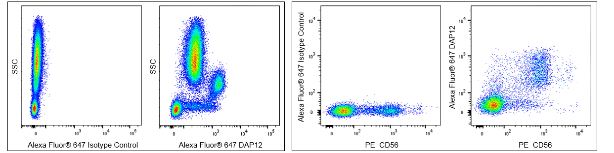
  6. Alexa Fluor® 647 fluorochrome emission is collected at the same instrument settings as for allophycocyanin (APC).
  7. For fluorochrome spectra and suitable instrument settings, please refer to our Multicolor Flow Cytometry web page at www.bdbiosciences.com/colors.
  8. Please refer to www.bdbiosciences.com/us/s/resources for technical protocols.
566603 Rev. 1
Antibody Details
Down Arrow Up Arrow
406288

The 406288 monoclonal antibody specifically recognizes DNAX-activation Protein 12 (DAP12) which is also known as Killer-activating receptor-associated protein (KARAP) or PLOSL. DAP12 is encoded by TYROBP (TYRO protein tyrosine kinase binding protein). The 12 kDa DAP12 protein contains a short extracellular domain, a transmembrane domain, and a cytoplasmic domain that contains a single immunoreceptor tyrosine-based activation motif (ITAM). DAP12 exists as a disulfide-linked homodimeric protein that is expressed by a variety of hematopoietic cells, including NK cells, monocytes, macrophages, dendritic cells, neutrophils, eosinophils, basophils, mast cells, and some NKT cells and γδ T cells. DAP12 functions as a signal transducing adaptor protein that associates with a variety of receptors involved in the detection of pathogens or host damage, including CD158 members of the Killer cell immunoglobulin like receptors (KIR) gene family, CD94-NKG2C and CD94-NKG2E heterodimeric receptors, Triggering receptor expressed on monocytes 1 (TREM-1) and TREM-2, and the NKp44 (CD336) receptor. These associations are mediated by intramembranal ionic attraction between a DAP12 aspartic acid residue and a lysine residue in the cognate receptor. The association between these receptors leads to either cellular activation or inhibition through dual tyrosine phosphorylation of the ITAM sequence by Src family kinases and further downstream signaling.


566603 Rev. 1
Format Details
Down Arrow Up Arrow
Alexa Fluor™ 647
Alexa Fluor™ 647 Dye is part of the BD red family of dyes. This is a small organic fluorochrome with an excitation maximum (Ex Max) at 653-nm and an emission maximum (Em Max) at 669-nm. Alexa Fluor 647 is designed to be excited by the Red laser (627-640 nm) and detected using an optical filter centered near 520-nm (e.g., a 660/20 nm bandpass filter). Please ensure that your instrument’s configurations (lasers and optical filters) are appropriate for this dye.
Alexa Fluor™ 647
Red 627-640 nm
653 nm
669 nm
566603 Rev.1
Citations & References
Down Arrow Up Arrow

Development References (3)

  1. Hamerman JA, Ni M, Killebrew JR, Chu CL, Lowell CA. The expanding roles of ITAM adapters FcRgamma and DAP12 in myeloid cells.. Immunol Rev. 2009; 232(1):42-58. (Biology). View Reference
  2. Lanier LL, Corliss BC, Wu J, Leong C, Phillips JH. Immunoreceptor DAP12 bearing a tyrosine-based activation motif is involved in activating NK cells.. Nature. 1998; 391(6668):703-7. (Biology). View Reference
  3. Lanier LL. DAP10- and DAP12-associated receptors in innate immunity.. Immunol Rev. 2009; 227(1):150-60. (Biology). View Reference
566603 Rev. 1

Please refer to Support Documents for Quality Certificates


Global - Refer to manufacturer's instructions for use and related User Manuals and Technical data sheets before using this products as described


Comparisons, where applicable, are made against older BD Technology, manual methods or are general performance claims.  Comparisons are not made against non-BD technologies, unless otherwise noted.

For Research Use Only. Not for use in diagnostic or therapeutic procedures.