Skip to main content Skip to navigation
PE Mouse Anti-Human CD27
Alert icon
Consider BD Horizon RealYellow™ 586 (RY586) Reagents, a bright and clean fluorochrome alternative to PE off the yellow-green laser. RY586 can be used alongside PE on spectral cytometers. More Info #
PE Mouse Anti-Human CD27
Multiparameter flow cytometric analysis of CD27 expression on human peripheral blood leucocyte populations. Human whole blood was stained with either PE Mouse IgG1, κ Isotype Control (Cat. No. 554680; Left Plot) or PE Mouse Anti-Human CD27 antibody (Cat. No. 566944/566945 Right Plot). Erythrocytes were lysed with BD Pharm Lyse™ Lysing Buffer (Cat. No. 555899). A two-parameter pseudocolor density plot showing the correlated expression of CD27 (or Ig Isotype control staining) versus side-light scatter (SSC-A) signals was derived from gated events with the forward and side-light scatter characteristics of intact leucocyte populations. Flow cytometric analysis was performed using a BD FACSCelesta™ Flow Cytometer System and FlowJo™ software. Data shown on this Technical Data Sheet are not lot specific.
Product Details
Down Arrow Up Arrow


BD Pharmingen™
CD27 antigen; TNFRSF7; S152; T14; Tp55
Human (QC Testing)
Mouse IgG1, κ
Not Reported
Flow cytometry (Routinely Tested)
5 µl
IV T-186
AB_2869961
Aqueous buffered solution containing BSA and ≤0.09% sodium azide.
RUO


Preparation And Storage

Store undiluted at 4°C and protected from prolonged exposure to light. Do not freeze. The monoclonal antibody was purified from tissue culture supernatant or ascites by affinity chromatography. The antibody was conjugated to the dye under optimum conditions and unconjugated antibody and free dye were removed.

Recommended Assay Procedures

BD™ CompBeads can be used as surrogates to assess fluorescence spillover (Compensation).  When fluorochrome conjugated antibodies are bound to CompBeads, they have spectral properties very similar to cells.   However, for some fluorochromes there can be small differences in spectral emissions compared to cells, resulting in spillover values that differ when compared to biological controls.  It is strongly recommended that when using a reagent for the first time, users compare the spillover on cell and CompBead to ensure that BD Comp beads are appropriate for your specific cellular application.

Product Notices

  1. This reagent has been pre-diluted for use at the recommended Volume per Test. We typically use 1 × 10^6 cells in a 100-µl experimental sample (a test).
  2. An isotype control should be used at the same concentration as the antibody of interest.
  3. Caution: Sodium azide yields highly toxic hydrazoic acid under acidic conditions. Dilute azide compounds in running water before discarding to avoid accumulation of potentially explosive deposits in plumbing.
  4. Source of all serum proteins is from USDA inspected abattoirs located in the United States.
  5. For fluorochrome spectra and suitable instrument settings, please refer to our Multicolor Flow Cytometry web page at www.bdbiosciences.com/colors.
  6. Please refer to http://regdocs.bd.com to access safety data sheets (SDS).
  7. Please refer to www.bdbiosciences.com/us/s/resources for technical protocols.
566945 Rev. 2
Antibody Details
Down Arrow Up Arrow
O323

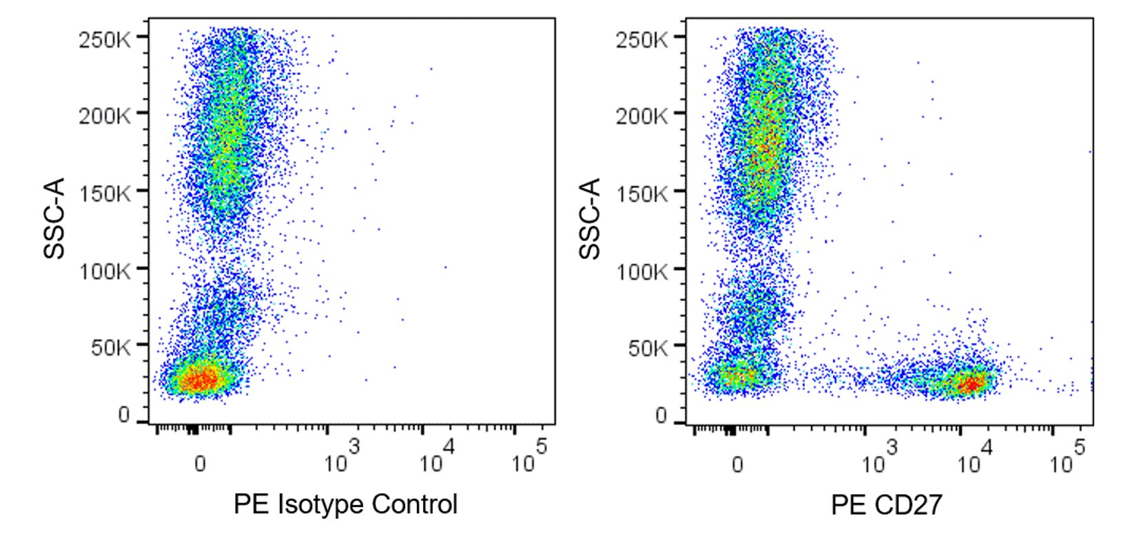
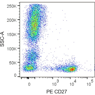
The O323 monoclonal antibody specifically recognizes CD27 which is also known as Tumor necrosis factor receptor superfamily member 7 (TNFRSF7), T14, Tp55, or S152. CD27 exists as a ~110-120 kDa disulfide-linked homodimer comprised of two single-pass type I transmembrane glycoproteins that are encoded by CD27 (CD27 molecule). CD27 is expressed on medullary thymocytes and T cells, with higher expression on activated T cells, and subsets of mature B cells and natural killer (NK) cells. A soluble 28-32 kDa form of CD27 is produced by lymphocytes upon cellular activation. Binding of the CD27 antigen, expressed on T cells, to its ligand, CD70 (CD27L), provides a costimulatory signal, leading to T cell proliferation, production of cytotoxic T cells, and enhanced production of cytokines. Binding of CD70 to CD27 expressed on B cells leads to B cell proliferation and the generation of plasma cells and immunoglobulin production. The CD27 antigen becomes hyperphosphorylated on serine residues upon activation of T cells. Signaling through the CD27 antigen activates NFκB and stress activated protein kinase (SAPK)/c Jun N terminal kinase (JNK).


566945 Rev. 2
Format Details
Down Arrow Up Arrow
PE
R-Phycoerythrin (PE), is part of the BD family of Phycobiliprotein dyes. This fluorochrome is a multimeric fluorescent phycobiliprotein with excitation maximum (Ex Max) of 496 nm and 566 nm and an emission maximum (Em Max) at 576 nm. PE is designed to be excited by the Blue (488 nm), Green (532 nm) and Yellow-Green (561 nm) lasers and detected using an optical filter centered near 575 nm (e.g., a 575/26-nm bandpass filter). As PE is excited by multiple lasers, this can result in cross-laser excitation and fluorescence spillover on instruments with various combinations of Blue, Green, and Yellow-Green lasers. Please ensure that your instrument’s configurations (lasers and optical filters) are appropriate for this dye.
altImg
PE
Yellow-Green 488 nm, 532 nm, 561 nm
496 nm, 566 nm
576 nm
566945 Rev.2
Citations & References
Down Arrow Up Arrow

Development References (5)

  1. Björkström NK, Béziat V, Cichocki F, et al. CD8 T cells express randomly selected KIRs with distinct specificities compared with NK cells.. Blood. 2012; 120(17):3455-65. (Clone-specific: Flow cytometry). View Reference
  2. Borst J, Hendriks J, Xiao Y. CD27 and CD70 in T cell and B cell activation.. Curr Opin Immunol. 2005; 17(3):275-81. (Biology). View Reference
  3. Klein U, Rajewsky K, Küppers R. Human immunoglobulin (Ig)M+IgD+ peripheral blood B cells expressing the CD27 cell surface antigen carry somatically mutated variable region genes: CD27 as a general marker for somatically mutated (memory) B cells.. J Exp Med. 1998; 188(9):1679-89. (Biology). View Reference
  4. Reiter C. T9. Cluster report: CD27. In: Knapp W. W. Knapp .. et al., ed. Leucocyte typing IV : white cell differentiation antigens. Oxford New York: Oxford University Press; 1989:350.
  5. Zola H. Leukocyte and stromal cell molecules : the CD markers. Hoboken, N.J.: Wiley-Liss; 2007.
View All (5) View Less
566945 Rev. 2

Please refer to Support Documents for Quality Certificates


Global - Refer to manufacturer's instructions for use and related User Manuals and Technical data sheets before using this products as described


Comparisons, where applicable, are made against older BD Technology, manual methods or are general performance claims.  Comparisons are not made against non-BD technologies, unless otherwise noted.

For Research Use Only. Not for use in diagnostic or therapeutic procedures.