Skip to main content Skip to navigation
BUV805 Mouse Anti-Human CD14
Alert icon
This product is the replacement for [565779].
BUV805 Mouse Anti-Human CD14
Multiparameter flow cytometric analysis of CD14 expression on human peripheral blood leucocytes. Whole blood was stained with either BD Horizon™ BUV805 Mouse IgG2a, κ Isotype Control (Cat. No. 612904; Left Plot) or BD Horizon BUV805 Mouse Anti-Human CD14 antibody (Cat. No. 612902/612903; Right Plot). Erythrocytes were lysed with BD FACS™ Lysing Solution (Cat. No. 349202). The two-parameter flow cytometric contour plot showing the correlated expression of CD14 (or Ig Isotype control staining) versus side-light scatter (SSC-A) signals were derived from gated events with the forward and side-light scatter characteristics of intact leucocyte populations. Flow cytometric analysis was performed using a BD™ LSR II Flow Cytometer System.
Product Details
Down Arrow Up Arrow


BD Horizon™
LPS receptor; LPS-R; Myeloid cell-specific leucine-rich glycoprotein
Human (QC Testing), Rhesus, Cynomolgus, Baboon, Dog (Tested in Development)
Mouse IgG2a, κ
Human CD14 Protein
Flow cytometry (Routinely Tested)
5 µl
II M34; III M329
929
AB_2870189
Aqueous buffered solution containing ≤0.09% sodium azide.
RUO


Preparation And Storage

Store undiluted at 4°C and protected from prolonged exposure to light. Do not freeze. The monoclonal antibody was purified from tissue culture supernatant or ascites by affinity chromatography. The antibody was conjugated with BD Horizon BUV805 under optimum conditions, and unconjugated antibody and free BD Horizon BUV805 were removed.

Recommended Assay Procedures

BD™ CompBeads can be used as surrogates to assess fluorescence spillover (Compensation). When fluorochrome-conjugated antibodies are bound to BD CompBeads, they have spectral properties very similar to cells. However, for some fluorochromes there can be small differences in spectral emissions compared to cells, resulting in spillover values that differ when compared to biological controls. It is strongly recommended that when using a reagent for the first time, users compare the spillover on cells and BD CompBeads. This will ensure that BD CompBeads are appropriate for your specific cellular application.

For optimal and reproducible results, BD Horizon Brilliant Stain Buffer should be used anytime two or more BD Horizon Brilliant dyes are used in the same experiment.  Fluorescent dye interactions may cause staining artifacts which may affect data interpretation.  The BD Horizon Brilliant Stain Buffer was designed to minimize these interactions.  More information can be found in the Technical Data Sheet of the BD Horizon Brilliant Stain Buffer (Cat. No. 563794/566349) or the BD Horizon Brilliant Stain Buffer Plus (Cat. No. 566385).

Note:  When using high concentrations of antibody, background binding of this dye to erythroid cell subsets (mature erythrocytes and precursors) has been observed.  For researchers studying these cell populations, or in cases where light scatter gating does not adequately exclude these cells from the analysis, this background may be an important factor to consider when selecting reagents for panel(s).

Product Notices

  1. This reagent has been pre-diluted for use at the recommended Volume per Test. We typically use 1 × 10^6 cells in a 100-µl experimental sample (a test).
  2. An isotype control should be used at the same concentration as the antibody of interest.
  3. Caution: Sodium azide yields highly toxic hydrazoic acid under acidic conditions. Dilute azide compounds in running water before discarding to avoid accumulation of potentially explosive deposits in plumbing.
  4. For fluorochrome spectra and suitable instrument settings, please refer to our Multicolor Flow Cytometry web page at www.bdbiosciences.com/colors.
  5. BD Horizon Brilliant Ultraviolet 805 is covered by one or more of the following US patents: 8,110,673, 8,158,444; 8,227,187; 8,575,303; 8,354,239.
  6. BD Horizon Brilliant Stain Buffer is covered by one or more of the following US patents: 8,110,673; 8,158,444; 8,575,303; 8,354,239.
  7. Species cross-reactivity detected in product development may not have been confirmed on every format and/or application.
  8. Please refer to www.bdbiosciences.com/us/s/resources for technical protocols.
612902 Rev. 2
Antibody Details
Down Arrow Up Arrow
M5E2

The M5E2 monoclonal antibody specifically binds to CD14, a 53–55 kDa glycosylphosphatidylinositol (GPI)-anchored single chain glycoprotein expressed at high levels on monocytes. Additionally, the anti-CD14 antibody reacts with interfollicular macrophages, reticular dendritic cells, and some Langerhans cells. CD14 has been identified as a high affinity cell-surface receptor for complexes of lipopolysaccharide (LPS) and serum LPS-binding protein, LPB.

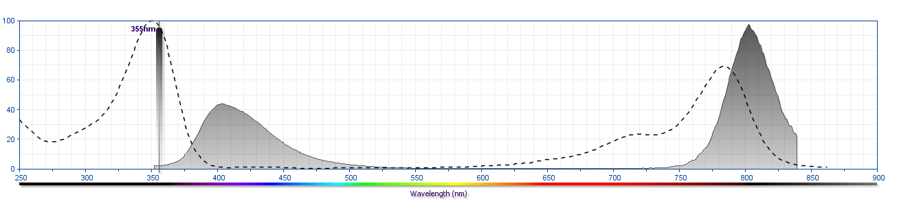
The antibody was conjugated to BD Horizon BUV805 which is part of the BD Horizon Brilliant™ Ultraviolet family of dyes. This dye is a tandem fluorochrome with an Ex Max near 350 nm and an Em Max near 805 nm. BD Horizon Brilliant BUV805 can be excited by the ultraviolet laser (355 nm) and detected with a 820/60 nm filter and a 770 nm LP.

612902 Rev. 2
Format Details
Down Arrow Up Arrow
BUV805
The BD Horizon Brilliant™ Ultraviolet 805 (BUV805) Dye is part of the BD Horizon Brilliant™ Ultraviolet family of dyes. This tandem fluorochrome is comprised of a BUV395 donor with an excitation maximum (Ex Max) of 351-nm and an acceptor dye with an emission maximum (Em Max) at 803-nm. BUV805, driven by BD innovation, is designed to be excited by the ultraviolet laser (355 nm) and detected using an optical filter centered near 805-nm (e.g., a 820/60 or a 780/60 bandpass filter). Please ensure that your instrument’s configurations (lasers and optical filters) are appropriate for this dye.
altImg
BUV805
Ultraviolet 355 nm
351 nm
803 nm
612902 Rev.2
Citations & References
Down Arrow Up Arrow

Development References (7)

  1. Bernstein ID, Self S. Joint report of the Myeloid Section of the Second International Workshop on Human Leukocyte Differentiation Antigens. In: Reinherz EL, Haynes BF, Nadler LM, Bernstein ID, ed. Leukocyte Typing II: Human Myeloid and Hematopoietic Cells. New York, NY: Springer-Verlag; 1986:1-25.
  2. Elliott L, Brooks W, Roszman T. Inhibition of anti-CD3 monoclonal antibody-induced T-cell proliferation by dexamethasone, isoproterenol, or prostaglandin E2 either alone or in combination.. Cell Mol Neurobiol. 1992; 12(5):411-27. (Clone-specific: Cell separation). View Reference
  3. Knapp W. W. Knapp .. et al., ed. Leucocyte typing IV : white cell differentiation antigens. Oxford New York: Oxford University Press; 1989:1-1182.
  4. Lin G-X, Yang X, Hollemweguer E, et al. Cross-reactivity of CD antibodies in eight animal species. In: Mason D. David Mason .. et al., ed. Leucocyte typing VII : white cell differentiation antigens : proceedings of the Seventh International Workshop and Conference held in Harrogate, United Kingdom. Oxford: Oxford University Press; 2002:519-523.
  5. McMichael AJ. A.J. McMichael .. et al., ed. Leucocyte typing III : white cell differentiation antigens. Oxford New York: Oxford University Press; 1987:1-1050.
  6. Schlossman SF. Stuart F. Schlossman .. et al., ed. Leucocyte typing V : white cell differentiation antigens : proceedings of the fifth international workshop and conference held in Boston, USA, 3-7 November, 1993. Oxford: Oxford University Press; 1995.
  7. Wright SD, Ramos RA, Tobias PS, Ulevitch RJ, Mathison JC. CD14, a receptor for complexes of lipopolysaccharide (LPS) and LPS binding protein. Science. 1990; 249(4975):1431-1433. (Biology). View Reference
View All (7) View Less
612902 Rev. 2

Please refer to Support Documents for Quality Certificates


Global - Refer to manufacturer's instructions for use and related User Manuals and Technical data sheets before using this products as described


Comparisons, where applicable, are made against older BD Technology, manual methods or are general performance claims.  Comparisons are not made against non-BD technologies, unless otherwise noted.

For Research Use Only. Not for use in diagnostic or therapeutic procedures.