APC-R700 Mouse Anti-Human CD69
Clone FN50 (also known as FN 50) (RUO)
- Brand BD Horizon™
- Alternative Name AIM; CLEC2C; EA1; GP32/28; Leu23; MLR-3; VEA; BL-AC/P26
- Vol. Per Test 5 µl
- Isotype Mouse IgG1, κ
- Reactivity Human (QC Testing) Rhesus, Cynomolgus, Baboon (Tested in Development)
- Application
Flow cytometry (Routinely Tested)
- Immunogen Anti-µ stimulated human B lymphocytes
- Workshop No. IV A91 (A091)
- Entrez Gene ID 969
- Storage Buffer Aqueous buffered solution containing BSA, protein stabilizer, glycerol and ≤0.09% sodium azide.
- Regulatory Status RUO
Regulatory Status Legend
Description
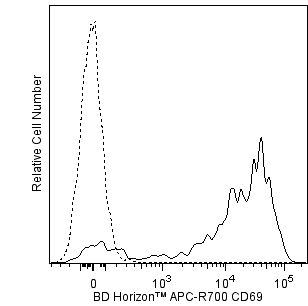
The FN50 monoclonal antibody specifically binds to human CD69. CD69 is also known as activation-induced molecule (AIM), early activation antigen (EA-1), very early activation antigen (VEA), C-type lectin domain family 2 member C (CLEC2C), MLR-3, GP32/28 and Leu-23. CD69 is a transmembrane type II homodimer receptor. CD69 is comprised of disulfide-linked, differentially glycosylated core protein subunits that are approximately 28 and 34 kDa in size. Each subunit contains a C-type lectin domain. CD69 is expressed on activated T, B, and natural killer (NK) lymphocytes, thymocytes, neutrophils, eosinophils and platelets. In normal peripheral blood, a small and variable percentage of lymphocytes typically express detectable membrane CD69 antigen. Upon activation, CD69 antigen expression increases on lymphocytes. Peak CD69 expression generally occurs within 18 hours of activation, preceding the appearance of HLA-DR, IL-2Rα (CD25) and transferrin receptor (CD71). CD69 is highly expressed on the bright CD3+ subset of thymocytes. FN50 monoclonal antibody labels NK cells and most lymphocytes of the follicular mantle and perifollicular/interfollicular zone as well as germinal center T cells of lymph nodes and tonsils. Studies indicate that CD69 serves as a signaling receptor in the activation of a variety of cell types.
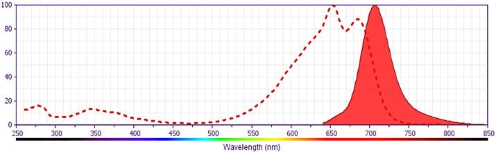
This antibody was conjugated to BD Horizon APC-R700, which has been developed exclusively by BD Biosciences as a better alternative to Alexa Fluor® 700. APC-R700 excites and emits at similar wavelengths to Alexa Fluor® 700 yet exhibits significantly improved brightness. This dye can be excited by the red laser and detected with the same filter set as Alexa Fluor® (eg, 730/45-nm filter).
Format
APC-R700 is a tandem fluorochrome that combines APC with R700, a proprietary organic dye. This dye has been developed exclusively by BD Biosciences as a brighter alternative to Alexa Fluor® 700. Due to similar excitation and emission properties, APC-R700 and Alexa Fluor® 700 cannot be used simultaneously.
Suggested Companion Products
Stain Buffer (FBS) RUO
500 mL
Cat No: 554656
Stain Buffer (BSA) RUO
500 mL
Cat No: 554657
APC-R700 Mouse Anti-Human CD69 FN50 (also known as FN 50) RUO
25 Tests
Cat No: 565155
Resources & Tools | ||||||
---|---|---|---|---|---|---|
Spectrum Viewer | Panel Designer | Spectrum Viewer | Download MSDS | Download TDS |
Preparation and Storage
Store undiluted at 4°C and protected from prolonged exposure to light. Do not freeze.
The monoclonal antibody was purified from tissue culture supernatant or ascites by affinity chromatography.
The antibody was conjugated with BD Horizon APC-R700 under optimum conditions, and unconjugated antibody and free BD Horizon APC-R700 were removed.
Product Notices
- This reagent has been pre-diluted for use at the recommended Volume per Test. We typically use 1 × 10^6 cells in a 100-µl experimental sample (a test).
- Please refer to www.bdbiosciences.com/us/s/resources for technical protocols.
- Alexa Fluor® is a registered trademark of Molecular Probes, Inc., Eugene, OR.
- Caution: Sodium azide yields highly toxic hydrazoic acid under acidic conditions. Dilute azide compounds in running water before discarding to avoid accumulation of potentially explosive deposits in plumbing.
- For fluorochrome spectra and suitable instrument settings, please refer to our Multicolor Flow Cytometry web page at www.bdbiosciences.com/colors.
- Source of all serum proteins is from USDA inspected abattoirs located in the United States.
- Species cross-reactivity detected in product development may not have been confirmed on every format and/or application.
- An isotype control should be used at the same concentration as the antibody of interest.