Skip to main content Skip to navigation
APC-R700 Mouse Anti-Human CD3
Alert icon
Consider BD Horizon™ Red 718 Reagents, bright small molecule alternative to APC-R700 with lower spread into the APC detector. More Info #
APC-R700 Mouse Anti-Human CD3
Flow cytometric analysis of CD3 expression on human peripheral blood lymphocytes. Human whole blood was stained with either BD Horizon™ APC-R700 Mouse IgG1, κ Isotype Control (Cat. No. 564974; dashed line histogram) or BD Horizon APC-R700 Mouse Anti-Human CD3 antibody (Cat. No. 565119/565120; solid line histogram). The erythrocytes were lysed with BD FACS™ Lysing Solution (Cat. No. 349202). The fluorescence histogram showing CD3 expression (or Ig Isotype control staining) was derived from events with the forward and side light-scatter characteristics of intact lymphocytes. Flow cytometric analysis was performed using a BD™ LSR II Flow Cytometry System.
Product Details
Down Arrow Up Arrow


BD Horizon™
CD3e; CD3E; T3E; TCRE; T-cell surface antigen T3/Leu-4 epsilon
Human (QC Testing)
Mouse BALB/c IgG1, κ
Human infant thymocytes and peripheral blood lymphocytes from a Sézary Syndrome donor
Flow cytometry (Routinely Tested)
5 µl
I WT3; III 471
916
AB_2744385
Aqueous buffered solution containing BSA, protein stabilizer, glycerol and ≤0.09% sodium azide.
RUO


Preparation And Storage

Store undiluted at 4°C and protected from prolonged exposure to light. Do not freeze. The monoclonal antibody was purified from tissue culture supernatant or ascites by affinity chromatography. The antibody was conjugated with BD Horizon APC-R700 under optimum conditions, and unconjugated antibody and free BD Horizon APC-R700 were removed.

Product Notices

  1. This reagent has been pre-diluted for use at the recommended Volume per Test. We typically use 1 × 10^6 cells in a 100-µl experimental sample (a test).
  2. Please refer to www.bdbiosciences.com/us/s/resources for technical protocols.
  3. Alexa Fluor® is a registered trademark of Molecular Probes, Inc., Eugene, OR.
  4. Caution: Sodium azide yields highly toxic hydrazoic acid under acidic conditions. Dilute azide compounds in running water before discarding to avoid accumulation of potentially explosive deposits in plumbing.
  5. For fluorochrome spectra and suitable instrument settings, please refer to our Multicolor Flow Cytometry web page at www.bdbiosciences.com/colors.
  6. Source of all serum proteins is from USDA inspected abattoirs located in the United States.
  7. An isotype control should be used at the same concentration as the antibody of interest.
565119 Rev. 1
Antibody Details
Down Arrow Up Arrow
UCHT1

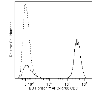
The UCHT1 monoclonal antibody specifically binds to the human CD3ε-chain, a 20-kDa subunit of the CD3/T cell antigen receptor complex. CD3ε is expressed on 70-80% of normal human peripheral blood lymphocytes and 60-85% of thymocytes. Studies from the HLDA Workshop show that this antibody is mitogenic for CD3ε-positive cells when used in conjunction with costimulatory agents such as pokeweed mitogen or anti-CD28 antibody. CD3 plays a central role in signal transduction during antigen recognition.  The UCHT1 antibody stains both surface and intracellular CD3ε unlike the other CD3 clone, HIT3a, that stains only extracellular CD3ε.

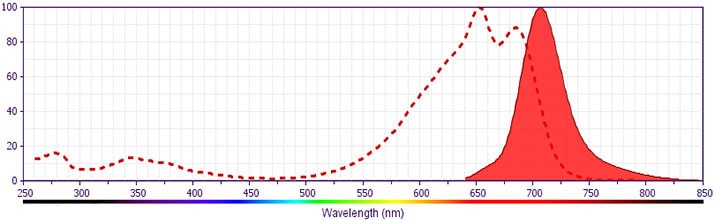
This antibody was conjugated to BD Horizon APC-R700, which has been developed exclusively by BD Biosciences as a better alternative to Alexa Fluor® 700. APC-R700 excites and emits at similar wavelengths to Alexa Fluor® 700 yet exhibits significantly improved brightness. This dye can be excited by the red laser and detected with the same filter set as Alexa Fluor® (eg, 730/45-nm filter).

565119 Rev. 1
Format Details
Down Arrow Up Arrow
APC-R700
The BD Horizon™ APC-R700 (APC-R700) Dye is a part of the BD APC red family of dyes. This tandem fluorochrome is comprised of an Allophycocyanin (APC) dye donor that has excitation maximum (Ex Max) of 651-nm and an acceptor dye, R700, with an emission maximum (Em Max) at 706-nm. APC-R700, driven by BD innovation, is designed to be excited by the red (627–640-nm) laser and detected using an optical filter centered near 710-nm (e.g., a 720/40-nm bandpass filter). APC-R700 is a brighter alternative to Alexa Fluor™ 700. Please ensure that your instrument’s configurations (lasers and optical filters) are appropriate for this dye.
altImg
APC-R700
Red 627-640 nm
651 nm
706 nm
565119 Rev.1
Citations & References
Down Arrow Up Arrow

Development References (6)

  1. Beverley PC, Callard RE. Distinctive functional characteristics of human "T" lymphocytes defined by E rosetting or a monoclonal anti-T cell antibody. Eur J Immunol. 1981; 11(4):329-334. (Immunogen: Flow cytometry, Fluorescence activated cell sorting). View Reference
  2. Burns GF, Boyd AW, Beverley PC. Two monoclonal anti-human T lymphocyte antibodies have similar biologic effects and recognize the same cell surface antigen. J Immunol. 1982; 129(4):1451-1457. (Clone-specific: Blocking, Functional assay, Immunoprecipitation, Inhibition, Radioimmunoassay). View Reference
  3. Ernst DN, Shih CC. CD3 complex. J Biol Regul Homeost Agents. 2000; 14(3):226-229. (Biology). View Reference
  4. Knapp W. W. Knapp .. et al., ed. Leucocyte typing IV : white cell differentiation antigens. Oxford New York: Oxford University Press; 1989:1-1182.
  5. McMichael AJ. A.J. McMichael .. et al., ed. Leucocyte typing III : white cell differentiation antigens. Oxford New York: Oxford University Press; 1987:1-1050.
  6. Van Wauwe JP, Goossens JG, Beverley PC. Human T lymphocyte activation by monoclonal antibodies; OKT3, but not UCHT1, triggers mitogenesis via an interleukin 2-dependent mechanism. J Immunol. 1984; 133(1):129-132. (Clone-specific: Flow cytometry, Functional assay, Stimulation). View Reference
View All (6) View Less
565119 Rev. 1

Please refer to Support Documents for Quality Certificates


Global - Refer to manufacturer's instructions for use and related User Manuals and Technical data sheets before using this products as described


Comparisons, where applicable, are made against older BD Technology, manual methods or are general performance claims.  Comparisons are not made against non-BD technologies, unless otherwise noted.

For Research Use Only. Not for use in diagnostic or therapeutic procedures.