
Preparation And Storage
Product Notices
- Since applications vary, each investigator should titrate the reagent to obtain optimal results.
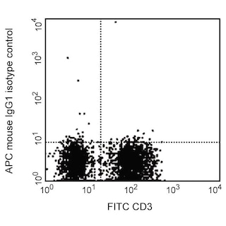
- An isotype control should be used at the same concentration as the antibody of interest.
- Caution: Sodium azide yields highly toxic hydrazoic acid under acidic conditions. Dilute azide compounds in running water before discarding to avoid accumulation of potentially explosive deposits in plumbing.
- This APC-conjugated reagent can be used in any flow cytometer equipped with a dye, HeNe, or red diode laser.
- For fluorochrome spectra and suitable instrument settings, please refer to our Multicolor Flow Cytometry web page at www.bdbiosciences.com/colors.
- Please refer to http://regdocs.bd.com to access safety data sheets (SDS).
- Please refer to www.bdbiosciences.com/us/s/resources for technical protocols.
Companion Products


The 3D6 monoclonal antibody specifically binds to c-Met (MET), which is also known as Hepatocyte growth factor receptor (HGFR) or Scatter factor receptor (SF receptor). c-Met is a 190 kDa single-pass type I transmembrane glycoprotein that is posttranslationally cleaved into a disulfide-linked extracellular α-chain and a transmembrane β-chain. c-Met functions as a receptor tyrosine kinase (RTK) that is normally involved in the development, regeneration, and survival of cells and tissues. c-Met is expressed on a variety of cell types including stem cells and progenitor cells, hepatocytes, keratinocytes, epithelial cells, endothelial cells, and neurons. c-Met is autophosphorylated when bound by its ligand, Hepatocyte Growth Factor (HGF). This leads to further activation of downstream signaling pathways that induce multiple responses including cellular migration, proliferation, survival, and angiogenesis. Abnormal c-Met expression or activity has been associated with tumorigenesis. The 3D6 antibody can reportedly function as an agonist by binding to and activating human but not mouse c-Met.

Development References (5)
-
Kong-Beltran M, Seshagiri S, Zha J, et al. Somatic mutations lead to an oncogenic deletion of met in lung cancer.. Cancer Res. 2006; 66(1):283-9. (Clone-specific: Activation, Bioassay, Functional assay, In vivo exacerbation, Stimulation). View Reference
-
Nguyen TH, Loux N, Dagher I, et al. Improved gene transfer selectivity to hepatocarcinoma cells by retrovirus vector displaying single-chain variable fragment antibody against c-Met.. Cancer Gene Ther. 2003; 10(11):840-9. (Clone-specific: Bioassay, Flow cytometry, Functional assay). View Reference
-
Ohashi K, Marion PL, Nakai H, et al. Sustained survival of human hepatocytes in mice: A model for in vivo infection with human hepatitis B and hepatitis delta viruses.. Nat Med. 2000; 6(3):327-31. (Immunogen: Activation, Functional assay, In vivo exacerbation, Stimulation). View Reference
-
Sakai K, Aoki S, Matsumoto K. Hepatocyte growth factor and Met in drug discovery.. J Biochem. 2015; 157(5):271-84. (Biology). View Reference
-
Wright TG, Singh VK, Li JJ, et al. Increased production and secretion of HGF alpha-chain and an antagonistic HGF fragment in a human breast cancer progression model.. Int J Cancer. 2009; 125(5):1004-15. (Clone-specific: ELISA). View Reference
Please refer to Support Documents for Quality Certificates
Global - Refer to manufacturer's instructions for use and related User Manuals and Technical data sheets before using this products as described
Comparisons, where applicable, are made against older BD Technology, manual methods or are general performance claims. Comparisons are not made against non-BD technologies, unless otherwise noted.
For Research Use Only. Not for use in diagnostic or therapeutic procedures.