


Preparation And Storage
Product Notices
- Since applications vary, each investigator should titrate the reagent to obtain optimal results.
- An isotype control should be used at the same concentration as the antibody of interest.
- Caution: Sodium azide yields highly toxic hydrazoic acid under acidic conditions. Dilute azide compounds in running water before discarding to avoid accumulation of potentially explosive deposits in plumbing.
- The Alexa Fluor®, Pacific Blue™, and Cascade Blue® dye antibody conjugates in this product are sold under license from Molecular Probes, Inc. for research use only, excluding use in combination with microarrays, or as analyte specific reagents. The Alexa Fluor® dyes (except for Alexa Fluor® 430), Pacific Blue™ dye, and Cascade Blue® dye are covered by pending and issued patents.
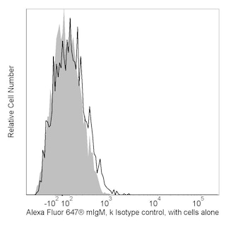
- Alexa Fluor® 647 fluorochrome emission is collected at the same instrument settings as for allophycocyanin (APC).
- Alexa Fluor® is a registered trademark of Molecular Probes, Inc., Eugene, OR.
- For fluorochrome spectra and suitable instrument settings, please refer to our Multicolor Flow Cytometry web page at www.bdbiosciences.com/colors.
- Please refer to www.bdbiosciences.com/us/s/resources for technical protocols.
Companion Products


The 48B3 monoclonal antibody specifically binds to CD167a which is also known as Discoidin domain receptor tyrosine kinase 1 (DDR1), Cell adhesion kinase (CAK), Mammary carcinoma kinase 10 (MCK10), or Neuroepithelial tyrosine kinase. CD167a is expressed on epithelial cells and at relatively low levels on immature dendritic cells and B lymphocytes. CD167a is a single-pass type I transmembrane glycoprotein. The extracellular CD167a discoidin homology domain mediates binding to various types of collagen. The binding of collagen leads to the activation of the cytoplasmic tyrosine kinase domain. The CD167a receptor tyrosine kinase (RTK) plays a role in regulating cellular proliferation, morphogenesis, differentiation, and collagen synthesis, as well as the synthesis of metalloproteases and extracellular matrix remodeling. CD167a may be involved in tumor invasion and metastasis. The 48B3 antibody does not recognize CD167b (DDR2).
Development References (4)
-
Bühring HJ, Buckley CD, Simmins DL. Review—CD167a—DDR1; discoidin domain receptor 1. In: Mason D. David Mason .. et al., ed. Leucocyte typing VII : white cell differentiation antigens : proceedings of the Seventh International Workshop and Conference held in Harrogate, United Kingdom. Oxford: Oxford University Press; 2002:21.
-
Johnson JD, Edman JC, Rutter WJ. A receptor tyrosine kinase found in breast carcinoma cells has an extracellular discoidin I-like domain.. Proc Natl Acad Sci USA. 1993; 90(22):10891. (Biology). View Reference
-
Vogel W, Gish GD, Alves F, Pawson T. The discoidin domain receptor tyrosine kinases are activated by collagen.. Mol Cell. 1997; 1(1):13-23. (Biology). View Reference
-
Zola H, Swart B, Boumsell L, Mason DY. Human Leucocyte Differentiation Antigen nomenclature: update on CD nomenclature. Report of IUIS/WHO Subcommittee.. J Immunol Methods. 2003; 275(1-2):1-8. (Clone-specific: Flow cytometry). View Reference
Please refer to Support Documents for Quality Certificates
Global - Refer to manufacturer's instructions for use and related User Manuals and Technical data sheets before using this products as described
Comparisons, where applicable, are made against older BD Technology, manual methods or are general performance claims. Comparisons are not made against non-BD technologies, unless otherwise noted.
For Research Use Only. Not for use in diagnostic or therapeutic procedures.