Skip to main content Skip to navigation
Alexa Fluor® 647 Mouse Anti-Human BCL7A
Flow cytometric analysis of BCL7A expression on human Burkitt's lymphoma cell lines. Daudi (Left Panel) and Raji (Right Panel) cells were fixed and permeabilized with BD Pharmingen™ Transcription Factor Buffer Set (Cat. No. 562725). The cells were stained with either Alexa Fluor® 647 Mouse IgG1, κ Isotype Control (Cat. No. 557732; 0.5 µg, dashed line histograms) or Alexa Fluor® 647 Mouse Anti-Human BCL7A (Cat. No. 566425; 0.5 µg, solid line histograms). The fluorescence histograms showing BCL7A expression (or Ig Isotype control staining), were derived from gated events with the forward and side light-scatter characteristics of viable cells. Flow cytometric analysis was performed using a BD LSRFortessa™ X-20 Flow Cytometer System. Data shown on this Technical Data Sheet are not lot specific.
Alexa Fluor® 647 Mouse Anti-Human BCL7A
Immunohistofluorescent staining of BCL7A in human tonsil. Following antigen retrieval with BD Pharmingen™ Retrievagen A Buffer (Cat. No. 550524), the sections of formalin-fixed, paraffin-embedded tissue were stained with BD Horizon™ BV421 Mouse Anti-Human CD45 (Cat. No. 563879, cytoplasmic staining, pseudocolored green) and Alexa Fluor® 647 Mouse Anti-BCL7A (Cat. No. 566425, nuclear staining, pseudocolored red) at 0.5 µg. Photography was performed on a Molecular Devices standard epifluorescence microscope. Original magnification, 20×.
Flow cytometric analysis of BCL7A expression on human Burkitt's lymphoma cell lines. Daudi (Left Panel) and Raji (Right Panel) cells were fixed and permeabilized with BD Pharmingen™ Transcription Factor Buffer Set (Cat. No. 562725). The cells were stained with either Alexa Fluor® 647 Mouse IgG1, κ Isotype Control (Cat. No. 557732; 0.5 µg, dashed line histograms) or Alexa Fluor® 647 Mouse Anti-Human BCL7A (Cat. No. 566425; 0.5 µg, solid line histograms). The fluorescence histograms showing BCL7A expression (or Ig Isotype control staining), were derived from gated events with the forward and side light-scatter characteristics of viable cells. Flow cytometric analysis was performed using a BD LSRFortessa™ X-20 Flow Cytometer System. Data shown on this Technical Data Sheet are not lot specific.
Immunohistofluorescent staining of BCL7A in human tonsil. Following antigen retrieval with BD Pharmingen™ Retrievagen A Buffer (Cat. No. 550524), the sections of formalin-fixed, paraffin-embedded tissue were stained with BD Horizon™ BV421 Mouse Anti-Human CD45 (Cat. No. 563879, cytoplasmic staining, pseudocolored green) and Alexa Fluor® 647 Mouse Anti-BCL7A (Cat. No. 566425, nuclear staining, pseudocolored red) at 0.5 µg. Photography was performed on a Molecular Devices standard epifluorescence microscope. Original magnification, 20×.
Product Details
Down Arrow Up Arrow


BD Pharmingen™
B-cell CLL/lymphoma 7 protein family member A; BCL7
Human (QC Testing)
Mouse BALB/c IgG1, κ
Human BCL7A Recombinant Protein
Intracellular staining (flow cytometry) (Routinely Tested), Immunofluorescence, Immunohistochemistry-paraffin (Tested During Development)
0.2 mg/ml
AB_2739725
Aqueous buffered solution containing protein stabilizer and ≤0.09% sodium azide.
RUO


Preparation And Storage

Store undiluted at 4°C and protected from prolonged exposure to light. Do not freeze. The monoclonal antibody was purified from tissue culture supernatant or ascites by affinity chromatography. The antibody was conjugated to Alexa Fluor® 647 under optimum conditions, and unreacted Alexa Fluor® 647 was removed.

Product Notices

  1. Since applications vary, each investigator should titrate the reagent to obtain optimal results.
  2. Please refer to www.bdbiosciences.com/us/s/resources for technical protocols.
  3. Caution: Sodium azide yields highly toxic hydrazoic acid under acidic conditions. Dilute azide compounds in running water before discarding to avoid accumulation of potentially explosive deposits in plumbing.
  4. For fluorochrome spectra and suitable instrument settings, please refer to our Multicolor Flow Cytometry web page at www.bdbiosciences.com/colors.
  5. The Alexa Fluor®, Pacific Blue™, and Cascade Blue® dye antibody conjugates in this product are sold under license from Molecular Probes, Inc. for research use only, excluding use in combination with microarrays, or as analyte specific reagents. The Alexa Fluor® dyes (except for Alexa Fluor® 430), Pacific Blue™ dye, and Cascade Blue® dye are covered by pending and issued patents.
  6. Alexa Fluor® is a registered trademark of Molecular Probes, Inc., Eugene, OR.
  7. Alexa Fluor® 647 fluorochrome emission is collected at the same instrument settings as for allophycocyanin (APC).
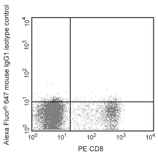
  8. An isotype control should be used at the same concentration as the antibody of interest.
566425 Rev. 1
Antibody Details
Down Arrow Up Arrow
15C

The 15C monoclonal antibody specifically recognizes the B-cell CLL/lymphoma 7A intranuclear protein (BCL7A). BCL7A is encoded by BCL7A (BCL tumor suppressor 7A) of the the BCL7 gene family. The three known BCL7 proteins (BCL7A, BCL7B, and BCL7C) share N-terminal sequence homology and may play a role in chromatin remodeling. BCL7A is normally expressed in germinal center (GC), some marginal zone (MZ) and interfollicular B lymphocytes, follicular and plasmacytoid dendritic cells, and cortical thymocytes, but not peripheral T lymphocytes. BCL7A has been associated with chromosomal aberrations leading to B-cell non-Hodgkin lymphoma, and is expressed in the majority of precursor and mature B cell lymphomas. BCL7A may be highly expressed in GC-related B-cell lymphoma but is not expressed in mature T-cell malignancies. The 15C antibody reportedly recognizes an epitope within the C-terminal region of BCL7A and has no cross-reactivity with human BCL7B or BCL7C.

566425 Rev. 1
Format Details
Down Arrow Up Arrow
Alexa Fluor™ 647
Alexa Fluor™ 647 Dye is part of the BD red family of dyes. This is a small organic fluorochrome with an excitation maximum (Ex Max) at 653-nm and an emission maximum (Em Max) at 669-nm. Alexa Fluor 647 is designed to be excited by the Red laser (627-640 nm) and detected using an optical filter centered near 520-nm (e.g., a 660/20 nm bandpass filter). Please ensure that your instrument’s configurations (lasers and optical filters) are appropriate for this dye.
Alexa Fluor™ 647
Red 627-640 nm
653 nm
669 nm
566425 Rev.1
Citations & References
Down Arrow Up Arrow

Development References (3)

  1. Jadayel DM, Osborne LR, Coignet LJ, et al. The BCL7 gene family: deletion of BCL7B in Williams syndrome. Gene. 1998; 224(1-2):35-44. (Biology).
  2. Morton LM, Purdue MP, Zheng T, et al. Risk of non-Hodgkin lymphoma associated with germline variation in genes that regulate the cell cycle, apoptosis, and lymphocyte development. Cancer Epidemiol Biomarkers Prev.. 2009; 18(4):1259-1270. (Biology).
  3. Ramos-Medina R, Montes-Moreno S, Maestre L, et al. Br J Haematol. 2013; 160(1):106-109. (Immunogen: Immunofluorescence, Immunohistochemistry, Western blot). View Reference
566425 Rev. 1

Please refer to Support Documents for Quality Certificates


Global - Refer to manufacturer's instructions for use and related User Manuals and Technical data sheets before using this products as described


Comparisons, where applicable, are made against older BD Technology, manual methods or are general performance claims.  Comparisons are not made against non-BD technologies, unless otherwise noted.

For Research Use Only. Not for use in diagnostic or therapeutic procedures.