Skip to main content Skip to navigation
Purified Mouse Anti-GFAP
Alert icon
This SKU will be discontinuing Apr 2024. For additional support, contact your local applications specialist. Contact Us #
Purified Mouse Anti-GFAP
Immunohistochemistry analysis of GFAP expression on human brain. Formalin-fixed, paraffin embedded section of human brain was stained with Purified Mouse Anti-GFAP (Cat. No. 556328) and visualized using a DAB chromogen and hematoxylin counterstain. Red arrow indicates an astrocyte (positive).
Purified Mouse Anti-GFAP
Flow cytometric analysis of GFAP expression on human brain cell line U373. U373 cells were stained with either Purified Mouse Anti-GFAP (solid line histogram) or Purified Mouse IgG2b, κ Isotype Control (Cat. No. 556654), followed by PE Goat Anti-Mouse Ig (Multiple Adsorption) (Cat. No. 550589). Fluorescent histograms were derived from gated events with the side and forward light-scattering characteristics of viable cells.
Immunohistochemistry analysis of GFAP expression on human brain. Formalin-fixed, paraffin embedded section of human brain was stained with Purified Mouse Anti-GFAP (Cat. No. 556328) and visualized using a DAB chromogen and hematoxylin counterstain. Red arrow indicates an astrocyte (positive).
Flow cytometric analysis of GFAP expression on human brain cell line U373. U373 cells were stained with either Purified Mouse Anti-GFAP (solid line histogram) or Purified Mouse IgG2b, κ Isotype Control (Cat. No. 556654), followed by PE Goat Anti-Mouse Ig (Multiple Adsorption) (Cat. No. 550589). Fluorescent histograms were derived from gated events with the side and forward light-scattering characteristics of viable cells.
Product Details
Down Arrow Up Arrow


BD Pharmingen™
Glial Fibrillary Acidic Protein, FLJ45472
Human (QC Testing), Rat (Tested in Development), Mouse,Pig,Dog,Chicken,Rabbit,Cow,Guinea Pig,Sheep (Reported)
Mouse IgG2b
Cow spinal cord homogenate
Flow cytometry (Routinely Tested), Immunofluorescence, Immunohistochemistry-formalin (antigen retrieval required) (Tested During Development), Immunohistochemistry-frozen, Western blot (Reported)
50 kDa
0.5 mg/ml
AB_396366
Aqueous buffered solution containing ≤0.09% sodium azide.
RUO


Preparation And Storage

Store undiluted at 4°C. The monoclonal antibody was purified from tissue culture supernatant or ascites by affinity chromatography.

Recommended Assay Procedures

BD Pharmingen offers additional GFAP specific antibodies: clone 2E1 (Cat. No. 556329); 4A11 (Cat. No. 556327); clones 1B4, 4A11, 2E1 combined and available as a "cocktail" (Cat. No. 556330). Applications include indirect immunofluorescence of tissue-cultured cells, immunohistochemical staining of formalin-fixed paraffin-embedded brain tissue sections (25 µg/ml); and western blot analysis (1-2 µg/ml). Rat brain is suggested as a positive control.

Product Notices

  1. Since applications vary, each investigator should titrate the reagent to obtain optimal results.
  2. An isotype control should be used at the same concentration as the antibody of interest.
  3. Caution: Sodium azide yields highly toxic hydrazoic acid under acidic conditions. Dilute azide compounds in running water before discarding to avoid accumulation of potentially explosive deposits in plumbing.
  4. Please refer to http://regdocs.bd.com to access safety data sheets (SDS).
  5. Sodium azide is a reversible inhibitor of oxidative metabolism; therefore, antibody preparations containing this preservative agent must not be used in cell cultures nor injected into animals. Sodium azide may be removed by washing stained cells or plate-bound antibody or dialyzing soluble antibody in sodium azide-free buffer. Since endotoxin may also affect the results of functional studies, we recommend the NA/LE (No Azide/Low Endotoxin) antibody format, if available, for in vitro and in vivo use.
  6. Please refer to www.bdbiosciences.com/us/s/resources for technical protocols.
556328 Rev. 12
Antibody Details
Down Arrow Up Arrow
1B4

GFAP (Glial Fibrillary Acid Protein) is the major protein of glial filaments in differentiated astrocytes. BD Pharmingen offers a panel of

monoclonal antibodies (4A11, 1B4, 2E1) that specifically recognize GFAP. They do not cross-react with other intermediate filaments such as

vimentin, neurofilament proteins, desmin, keratin, neurotubules or microfilaments. Bovine spinal cord homogenate was used as immunogen.

This antibody has broad species reactivity, recognizing GFAP in brain homogenates from human, mouse, rat, cow, sheep, dog, pig, rabbit,

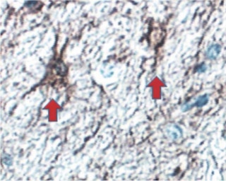
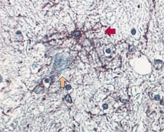
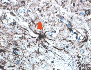
guinea pig and chicken. 1B4 is particularly useful for identifying GFAP in immunohistochemistry of frozen and formalin-fixed, paraffin-embedded brain tissue sections. Additional applications include western blot analysis and indirect immunofluorescence of tissue-cultured cells.

556328 Rev. 12
Format Details
Down Arrow Up Arrow
Purified
Tissue culture supernatant is purified by either protein A/G or affinity purification methods. Both methods yield antibody in solution that is free of most other soluble proteins, lipids, etc. This format provides pure antibody that is suitable for a number of downstream applications including: secondary labeling for flow cytometry or microscopy, ELISA, Western blot, etc.
Purified
556328 Rev.12
Citations & References
Down Arrow Up Arrow

Development References (3)

  1. McLendon RE, Bigner DD. Immunohistochemistry of the glial fibrillary acidic protein: basic and applied considerations. Brain Pathol. 1994; 4(3):221-228. (Clone-specific). View Reference
  2. McLendon RE, Burger PC, Pegram CN, Eng LF, Bigner DD. The immunohistochemical application of three anti-GFAP monoclonal antibodies to formalin-fixed, paraffin-embedded, normal and neoplastic brain tissues. J Neuropathol Exp Neurol. 1986; 45(6):692-703. (Clone-specific: Immunohistochemistry). View Reference
  3. Pegram CN, Eng LF, Wikstrand CJ, McComb RD, Lee YL, Bigner DD. Monoclonal antibodies reactive with epitopes restricted to glial fibrillary acidic proteins of several species. Neurochem Pathol. 1985; 3(2):119-138. (Clone-specific: Immunohistochemistry, Western blot). View Reference
556328 Rev. 12

Please refer to Support Documents for Quality Certificates


Global - Refer to manufacturer's instructions for use and related User Manuals and Technical data sheets before using this products as described


Comparisons, where applicable, are made against older BD Technology, manual methods or are general performance claims.  Comparisons are not made against non-BD technologies, unless otherwise noted.

For Research Use Only. Not for use in diagnostic or therapeutic procedures.