Skip to main content Skip to navigation
Alexa Fluor® 647 Rat Anti-Mouse CD155
Multicolor flow cytometric analysis of CD155 expression on mouse thymocytes. C57BL/6 mouse thymocytes were preincubated with Purified Rat Anti-Mouse CD16/CD32 antibody (Mouse BD Fc Block™) (Cat. No. 553141/553142). The cells were then stained with BD Horizon™ BV421 Rat Anti-Mouse CD4 (Cat. No. 562891) or PE Rat Anti-Mouse CD8a (Cat. No. 553033/553032/561095) antibodies, and either Alexa Fluor® 647 Rat IgG2a, κ Isotype Control (Cat. No. 557690; dashed line histograms) or Alexa Fluor® 647 Rat Anti-Mouse CD155 antibody (Cat. No. 566630; solid line histograms) at 0.5 µg/dose. BD Via-Probe™ Cell Viability 7-AAD Solution (Cat. No. 555815/555816) was added to cells right before analysis. The fluorescence histograms showing CD155 expression (or Ig Isotype control staining) were derived from CD4-CD8+ (Upper Left Plot), CD4+CD8+ (Upper Right), CD4-CD8- (Lower Left), or CD4+CD8- (Lower Right) gated events with the forward and side light-scatter characteristics of viable (7-AAD-negative) thymocytes. Flow cytometric analysis was performed using a BD LSRFortessa™ Cell Analyzer System. Data shown in this Technical Data Sheet are not lot specific.
Product Details
Down Arrow Up Arrow


BD Pharmingen™
Poliovirus receptor; Pvr; Tumor-associated antigen 1; Taa1; Tage4; HVED
Mouse (QC Testing)
Rat IgG2a, κ
Mouse CD155 Recombinant Protein
Flow cytometry (Routinely Tested)
0.2 mg/ml
52118
AB_2869798
Aqueous buffered solution containing ≤0.09% sodium azide.
RUO


Preparation And Storage

Store undiluted at 4°C and protected from prolonged exposure to light. Do not freeze. The monoclonal antibody was purified from tissue culture supernatant or ascites by affinity chromatography. The antibody was conjugated to Alexa Fluor® 647 under optimum conditions, and unreacted Alexa Fluor® 647 was removed.

Product Notices

  1. Since applications vary, each investigator should titrate the reagent to obtain optimal results.
  2. An isotype control should be used at the same concentration as the antibody of interest.
  3. Caution: Sodium azide yields highly toxic hydrazoic acid under acidic conditions. Dilute azide compounds in running water before discarding to avoid accumulation of potentially explosive deposits in plumbing.
  4. The Alexa Fluor®, Pacific Blue™, and Cascade Blue® dye antibody conjugates in this product are sold under license from Molecular Probes, Inc. for research use only, excluding use in combination with microarrays, or as analyte specific reagents. The Alexa Fluor® dyes (except for Alexa Fluor® 430), Pacific Blue™ dye, and Cascade Blue® dye are covered by pending and issued patents.
  5. Alexa Fluor® is a registered trademark of Molecular Probes, Inc., Eugene, OR.
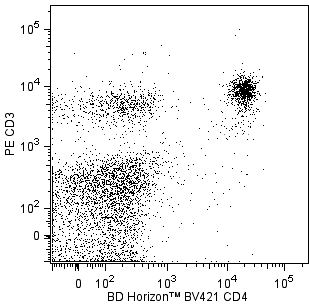
  6. Alexa Fluor® 647 fluorochrome emission is collected at the same instrument settings as for allophycocyanin (APC).
  7. For fluorochrome spectra and suitable instrument settings, please refer to our Multicolor Flow Cytometry web page at www.bdbiosciences.com/colors.
  8. Please refer to www.bdbiosciences.com/us/s/resources for technical protocols.
566630 Rev. 1
Antibody Details
Down Arrow Up Arrow
3F1

The 3F1 monoclonal antibody specifically binds to CD155, which is also known as Poliovirus receptor (Pvr) or Tumor-associated antigen 1 (Taa1). CD155 is a type I transmembrane glycoprotein that belongs to the Ig supergene family. CD155 is an adhesion receptor that binds to different ligands including nectin-3, CD96, CD226, TIGIT, and the extracellular matrix protein vitronectin. It is highly expressed on double positive thymocytes and variably expressed on mature thymocytes and T cells, including regulatory T cells and NKT cells. CD155 is also differentially expressed on subsets of B cells, plasma cells, dendritic cells, and monocytes. CD155 expression is upregulated by activated T cells, B cells, and dendritic cells. CD155 is involved in forming adherens junctions between adjacent epithelial or endothelial cells. CD155 plays roles in regulating cell growth, adhesion, motility, migration, and cell-mediated cytotoxicity. CD155-deficient mice exhibit impaired secondary humoral immune responses to orally administered antigens.

566630 Rev. 1
Format Details
Down Arrow Up Arrow
Alexa Fluor™ 647
Alexa Fluor™ 647 Dye is part of the BD red family of dyes. This is a small organic fluorochrome with an excitation maximum (Ex Max) at 653-nm and an emission maximum (Em Max) at 669-nm. Alexa Fluor 647 is designed to be excited by the Red laser (627-640 nm) and detected using an optical filter centered near 520-nm (e.g., a 660/20 nm bandpass filter). Please ensure that your instrument’s configurations (lasers and optical filters) are appropriate for this dye.
Alexa Fluor™ 647
Red 627-640 nm
653 nm
669 nm
566630 Rev.1
Citations & References
Down Arrow Up Arrow

Development References (3)

  1. Danisch S, Qiu Q, Seth S, et al. CD226 interaction with CD155 impacts on retention and negative selection of CD8 positive thymocytes as well as T cell differentiation to follicular helper cells in Peyer's Patches.. Immunobiology. 2013; 218(2):152-8. (Biology). View Reference
  2. Maier MK, Seth S, Czeloth N, et al. The adhesion receptor CD155 determines the magnitude of humoral immune responses against orally ingested antigens.. Eur J Immunol. 2007; 37(8):2214-25. (Immunogen: ELISA, Flow cytometry). View Reference
  3. Stanietsky N, Rovis TL, Glasner A, et al. Mouse TIGIT inhibits NK-cell cytotoxicity upon interaction with PVR.. Eur J Immunol. 2013; 43(8):2138-50. (Clone-specific: Flow cytometry). View Reference
566630 Rev. 1

Please refer to Support Documents for Quality Certificates


Global - Refer to manufacturer's instructions for use and related User Manuals and Technical data sheets before using this products as described


Comparisons, where applicable, are made against older BD Technology, manual methods or are general performance claims.  Comparisons are not made against non-BD technologies, unless otherwise noted.

For Research Use Only. Not for use in diagnostic or therapeutic procedures.