Skip to main content Skip to navigation
Purified Mouse Anti-Human CD276
CD276 expression on human immature dendritic cells. Human PBMC were isolated using Ficoll-Paque density gradient medium and cultured for three hours. The non-adherent fraction was removed, and the remaining adherent cells were cultured with Recombinant Human IL-4 (Cat. No. 554605) and Recombinant Human GM-CSF (Cat. No. 550068) for five days. The cells were harvested and stained with either Purified Mouse Anti-Human CD276 (Cat. No. 565828, solid-line histogram) or Purified Mouse IgG1 κ Isotype Control (Cat. No. 554121, dashed-line histogram). After washing with one time with BD Pharmingen™ Stain Buffer (FBS) (Cat. No. 554656), the second-step reagent PE Goat Anti-Mouse Ig (Cat. No. 550589) was added. Samples were washed  with BD Pharmingen™ Stain Buffer (FBS) and data was acquired. The histograms were derived from gated events with the forward and side light-scatter characteristics of viable dendritic cells. Flow cytometry was performed using BD™ LSR II Fortessa Flow Cytometer System. Overlay plots were generated using FlowJo™ Software.
Purified Mouse Anti-Human CD276
Immunohistochemical staining of CD276 on human tonsil (Top Row) and placenta (Bottom Row). The acetone-fixed frozen sections were stained with either Purified Mouse IgG1 κ Isotype Control (Cat. No.550878; Left Column) or Purified Mouse Anti-Human CD276 (Cat. No. 565828; Right Column). A three-step staining procedure that employed Biotin Goat Anti-Mouse Ig (Cat. No. 550337), Streptavidin HRP (Cat. No. 550946), and the DAB Substrate Kit (Cat. No. 550880) was used to develop the primary staining reagents. Counterstaining was with Hematoxylin. Top Row: CD276 expression  can be observed on some B lymphocytes, dendritic cells and macrophages in the tonsil. Original magnification: 20×. Bottom Row: CD276 expression on trophoblast and Hofbauer cells can be observed in the placenta. Original magnification: 40×.
CD276 expression on human immature dendritic cells. Human PBMC were isolated using Ficoll-Paque density gradient medium and cultured for three hours. The non-adherent fraction was removed, and the remaining adherent cells were cultured with Recombinant Human IL-4 (Cat. No. 554605) and Recombinant Human GM-CSF (Cat. No. 550068) for five days. The cells were harvested and stained with either Purified Mouse Anti-Human CD276 (Cat. No. 565828, solid-line histogram) or Purified Mouse IgG1 κ Isotype Control (Cat. No. 554121, dashed-line histogram). After washing with one time with BD Pharmingen™ Stain Buffer (FBS) (Cat. No. 554656), the second-step reagent PE Goat Anti-Mouse Ig (Cat. No. 550589) was added. Samples were washed  with BD Pharmingen™ Stain Buffer (FBS) and data was acquired. The histograms were derived from gated events with the forward and side light-scatter characteristics of viable dendritic cells. Flow cytometry was performed using BD™ LSR II Fortessa Flow Cytometer System. Overlay plots were generated using FlowJo™ Software.
Immunohistochemical staining of CD276 on human tonsil (Top Row) and placenta (Bottom Row). The acetone-fixed frozen sections were stained with either Purified Mouse IgG1 κ Isotype Control (Cat. No.550878; Left Column) or Purified Mouse Anti-Human CD276 (Cat. No. 565828; Right Column). A three-step staining procedure that employed Biotin Goat Anti-Mouse Ig (Cat. No. 550337), Streptavidin HRP (Cat. No. 550946), and the DAB Substrate Kit (Cat. No. 550880) was used to develop the primary staining reagents. Counterstaining was with Hematoxylin. Top Row: CD276 expression  can be observed on some B lymphocytes, dendritic cells and macrophages in the tonsil. Original magnification: 20×. Bottom Row: CD276 expression on trophoblast and Hofbauer cells can be observed in the placenta. Original magnification: 40×.
Product Details
Down Arrow Up Arrow


BD Pharmingen™
B7-H3, B7H3, 4Ig-B7-H3, B7RP-2
Human (QC Testing)
Mouse BALB/c IgG1, κ
Human Monocyte-Derived Dendritic Cells
Flow cytometry (Routinely Tested), Immunohistochemistry-frozen (Tested During Development), Immunohistochemistry-paraffin (Not Recommended)
0.5 mg/ml
VIII 80650
AB_2739368
Aqueous buffered solution containing ≤0.09% sodium azide.
RUO


Preparation And Storage

Store undiluted at 4°C. The monoclonal antibody was purified from tissue culture supernatant or ascites by affinity chromatography.

Product Notices

  1. Since applications vary, each investigator should titrate the reagent to obtain optimal results.
  2. Please refer to www.bdbiosciences.com/us/s/resources for technical protocols.
  3. Caution: Sodium azide yields highly toxic hydrazoic acid under acidic conditions. Dilute azide compounds in running water before discarding to avoid accumulation of potentially explosive deposits in plumbing.
  4. Sodium azide is a reversible inhibitor of oxidative metabolism; therefore, antibody preparations containing this preservative agent must not be used in cell cultures nor injected into animals. Sodium azide may be removed by washing stained cells or plate-bound antibody or dialyzing soluble antibody in sodium azide-free buffer. Since endotoxin may also affect the results of functional studies, we recommend the NA/LE (No Azide/Low Endotoxin) antibody format, if available, for in vitro and in vivo use.
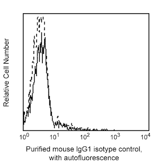
  5. An isotype control should be used at the same concentration as the antibody of interest.
  6. Ficoll-Paque is a trademark of Amersham Biosciences Limited.
  7. FlowJo is a trademark of Tree Star Inc.
565828 Rev. 1
Antibody Details
Down Arrow Up Arrow
7-517

The 7-517 monoclonal antibody specifically binds to CD276, also known as B7-H3 (B7 homolog 3). CD276 is a type I transmembrane glycoprotein and member of the B7 family of immunoregulatory proteins. Although the CD276 gene transcript is expressed in nearly all normal tissues, the protein is preferentially expressed in tumors, tumor cell lines, sinonasal epithelial cells, and extravillous trophoblast and Hofbauer cells of the placenta. CD276 protein expression can be induced by activation of T and B lymphocytes, natural killer (NK) cells and antigen-presenting cells. The roles of CD276 in the regulation of immune responses, especially to cancer, are under investigation.  

565828 Rev. 1
Format Details
Down Arrow Up Arrow
Purified
Tissue culture supernatant is purified by either protein A/G or affinity purification methods. Both methods yield antibody in solution that is free of most other soluble proteins, lipids, etc. This format provides pure antibody that is suitable for a number of downstream applications including: secondary labeling for flow cytometry or microscopy, ELISA, Western blot, etc.
Purified
565828 Rev.1
Citations & References
Down Arrow Up Arrow

Development References (6)

  1. Sallusto F, Lanzavecchia A. Efficient presentation of soluble antigen by cultured human dendritic cells is maintained by granulocyte/macrophage colony-stimulating factor plus interleukin 4 and downregulated by tumor necrosis factor alpha.. J Exp Med. 1994; 179(4):1109-18. (Methodology). View Reference
  2. Sharpe AH, Freeman GJ. The B7-CD28 superfamily.. Nat Rev Immunol. 2002; 2(2):116-26. (Biology). View Reference
  3. Steinberger P. B7-H3 ameliorates GVHD.. Blood. 2015; 125(21):3219-21. (Biology). View Reference
  4. Steinberger P1, Majdic O, Derdak SV, et al. Molecular characterization of human 4Ig-B7-H3, a member of the B7 family with four Ig-like domains.. J Immunol. 2004; 172(4):2352-2359. (Immunogen: Flow cytometry, Western blot). View Reference
  5. Sun J, Liu C, Gao L, et al. Correlation between B7-H3 expression and rheumatoid arthritis: A new polymorphism haplotype is associated with increased disease risk.. Clin Immunol. 2015; 159(1):23-32. (Biology). View Reference
  6. Sun M, Richards S, Prasad DV, Mai XM, Rudensky A, Dong C. Characterization of mouse and human B7-H3 genes.. J Immunol. 2002; 168(12):6294-7. (Biology). View Reference
View All (6) View Less
565828 Rev. 1

Please refer to Support Documents for Quality Certificates


Global - Refer to manufacturer's instructions for use and related User Manuals and Technical data sheets before using this products as described


Comparisons, where applicable, are made against older BD Technology, manual methods or are general performance claims.  Comparisons are not made against non-BD technologies, unless otherwise noted.

For Research Use Only. Not for use in diagnostic or therapeutic procedures.