-
Training
- Flow Cytometry Basic Training
-
Product-Based Training
- BD FACSDiscover™ S8 Cell Sorter Product Training
- Accuri C6 Plus Product-Based Training
- FACSAria Product Based Training
- FACSCanto Product-Based Training
- FACSLyric Product-Based Training
- FACSMelody Product-Based Training
- FACSymphony Product-Based Training
- HTS Product-Based Training
- LSRFortessa Product-Based Training
- Advanced Training
-
- BD FACSDiscover™ S8 Cell Sorter Product Training
- Accuri C6 Plus Product-Based Training
- FACSAria Product Based Training
- FACSCanto Product-Based Training
- FACSLyric Product-Based Training
- FACSMelody Product-Based Training
- FACSymphony Product-Based Training
- HTS Product-Based Training
- LSRFortessa Product-Based Training
- United States (English)
-
Change country/language
Old Browser
This page has been recently translated and is available in French now.
Looks like you're visiting us from {countryName}.
Would you like to stay on the current country site or be switched to your country?


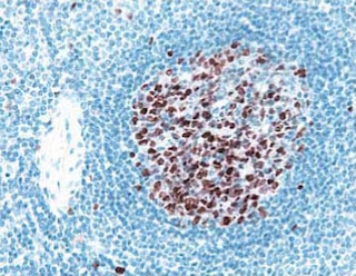
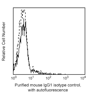
TOP ROW: Immunohistochemical analysis of Endosialin (CD248) in human tonsil. Following antigen retrieval with BD Retrievagen A Buffer (Cat. No. 550524), the formalin-fixed paraffin-embedded sections of human tomsil were stained with either Purified Mouse IgG1 κ Isotype Control (Cat. No.550878; Left Image) or Purified Mouse Anti-Human Endosialin (CD248) (Right Image). A three-step staining procedure that employs Biotin Goat Anti-Mouse Ig (Cat. No. 550337), Streptavidin HRP (Cat. No. 550946), and the DAB Substrate Kit (Cat. No. 550880) was used to develop the primary staining reagents. As shown in the right image, the CD248-specific antibody primarily stained stromal fibroblasts. Original magnification: 40×. BOTTOM LEFT: Flow cytometric analysis of human Endosialin (CD248) on human palatal mesenchyme cells. HEPM cells (ATTC, CRL-1486) were surface stained with either Purified Mouse IgG1, κ Isotype Control (Cat. No. 554121, dotted histogram) or Purified Mouse Anti-Human Endosialin (CD248) (solid line histogram). The second-step reagent was PE Goat Anti-Mouse Ig (Multiple Adsorption, Cat# 550589). The fluorescence histograms were derived from gated events with the forward and side light-scatter characteristics of viable cells. Flow cytometry was performed using a BD FACSCanto™ II Flow Cytometer System. BOTTOM RIGHT: Western blot analysis of human CD248 in human palatal mesenchyme cells. Lysates of HEPM cells (ATTC, CRL-1486) were prepared for electrophoresis (SDS-PAGE) in a 2D Tris-Glycine polyacrylamide gel. The proteins were transferred to PVDF membranes and then probed with 1 (lane 1), 0.5 (lane 2), and 0.25 (lane 3) µg/mL of Purified Mouse Anti-Human Endosialin (CD248). Specific staining was detected with HRP Goat Anti-Mouse Ig (Cat No 554002).


BD Pharmingen™ Purified Mouse Anti-Human Endosialin (CD248)

BD Pharmingen™ Purified Mouse Anti-Human Endosialin (CD248)

BD Pharmingen™ Purified Mouse Anti-Human Endosialin (CD248)

BD Pharmingen™ Purified Mouse Anti-Human Endosialin (CD248)

Regulatory Status Legend
Any use of products other than the permitted use without the express written authorization of Becton, Dickinson and Company is strictly prohibited.
Preparation And Storage
Product Notices
- Since applications vary, each investigator should titrate the reagent to obtain optimal results.
- Please refer to www.bdbiosciences.com/us/s/resources for technical protocols.
- An isotype control should be used at the same concentration as the antibody of interest.
- Caution: Sodium azide yields highly toxic hydrazoic acid under acidic conditions. Dilute azide compounds in running water before discarding to avoid accumulation of potentially explosive deposits in plumbing.
- Sodium azide is a reversible inhibitor of oxidative metabolism; therefore, antibody preparations containing this preservative agent must not be used in cell cultures nor injected into animals. Sodium azide may be removed by washing stained cells or plate-bound antibody or dialyzing soluble antibody in sodium azide-free buffer. Since endotoxin may also affect the results of functional studies, we recommend the NA/LE (No Azide/Low Endotoxin) antibody format, if available, for in vitro and in vivo use.
Companion Products


.png?imwidth=320)
The B1/35 monoclonal antibody specifically binds to CD248 which is also known as Endosialin, Tumor endothelial marker 1 (TEM1), and CD164 sialomucin-like 1 (CD164L1). CD248 is a heavily glycosylated, single-pass type I transmembrane protein. CD248 belongs to the Group XIV C-Type lectin family that includes CD93 and CD141/thrombomodulin. It is expressed on pericytes and stromal fibroblasts. Although the exact functions of CD248 remain to be determined, its expression is associated with angiogenesis and lymphoid tissue organization during development, as well as, with postnatal inflammation and tumor development and growth.
Development References (4)
-
Christian S, Ahorn H, Koehler A, et al. Molecular cloning and characterization of endosialin, a C-type lectin-like cell surface receptor of tumor endothelium. J Biol Chem. 2000; 276(10):7408-7414. (Biology). View Reference
-
MacFadyen JR, Haworth O, Roberston D, et al. Endosialin (TEM1, CD248) is a marker of stromal fibroblasts and is not selectively expressed on tumour endothelium. FEBS Lett. 2005; 579(12):2569-2575. (Immunogen: ELISA, Flow cytometry, Fluorescence microscopy, Immunoaffinity chromatography, Immunofluorescence, Immunoprecipitation). View Reference
-
Rettig WJ, Garin-Chesa P, Healey JH, Su SL, Jaffe EA, Old LJ. Identification of endosialin, a cell surface glycoprotein of vascular endothelial cells in human cancer. Proc Natl Acad Sci U S A. 1992; 89(22):10832-10836. (Biology). View Reference
-
St Croix B, Rago C, Velculescu V, et al. Genes expressed in human tumor endothelium. Science. 2000; 289(5482):1197-1202. (Biology). View Reference
Please refer to Support Documents for Quality Certificates
Global - Refer to manufacturer's instructions for use and related User Manuals and Technical data sheets before using this products as described
Comparisons, where applicable, are made against older BD Technology, manual methods or are general performance claims. Comparisons are not made against non-BD technologies, unless otherwise noted.
For Research Use Only. Not for use in diagnostic or therapeutic procedures.
Report a Site Issue
This form is intended to help us improve our website experience. For other support, please visit our Contact Us page.