-
Training
- Flow Cytometry Basic Training
-
Product-Based Training
- BD FACSDiscover™ S8 Cell Sorter Product Training
- Accuri C6 Plus Product-Based Training
- FACSAria Product Based Training
- FACSCanto Product-Based Training
- FACSLyric Product-Based Training
- FACSMelody Product-Based Training
- FACSymphony Product-Based Training
- HTS Product-Based Training
- LSRFortessa Product-Based Training
- Advanced Training
-
- BD FACSDiscover™ S8 Cell Sorter Product Training
- Accuri C6 Plus Product-Based Training
- FACSAria Product Based Training
- FACSCanto Product-Based Training
- FACSLyric Product-Based Training
- FACSMelody Product-Based Training
- FACSymphony Product-Based Training
- HTS Product-Based Training
- LSRFortessa Product-Based Training
- United States (English)
-
Change country/language
Old Browser
This page has been recently translated and is available in French now.
Looks like you're visiting us from {countryName}.
Would you like to stay on the current country site or be switched to your country?


.png)

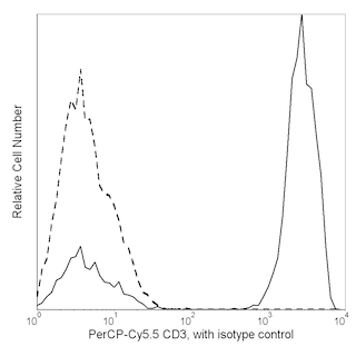
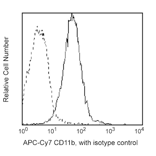
Two-color flow cytometric analysis of Fcμ Receptor expression on human peripheral blood mononuclear cells. PBMC were stained with PE Mouse Anti-Human Fcµ Receptor (Cat. No. 563017), PerCP-Cy™5.5 Mouse Anti-Human CD3 (560835), and APC-Cy™7 Mouse Anti-Human CD11b (557754/560914) antibodies. Two-color flow cytometric dot plots show the correlated expression patterns of CD3 (Left Panel) or CD11b (Right Panel) versus Fcμ Receptor for gated events with the forward and side light-scatter characteristics of viable leucocytes. Flow cytometry was performed using a BD™ LSR II Flow Cytometer System.
.png)

BD Pharmingen™ PE Mouse anti-Human Fcµ Receptor
.png)
Regulatory Status Legend
Any use of products other than the permitted use without the express written authorization of Becton, Dickinson and Company is strictly prohibited.
Preparation And Storage
Recommended Assay Procedures
Enhanced Fc µ Receptor staining intensity can be achieved by culturing cells overnight in complete media.
Product Notices
- This reagent has been pre-diluted for use at the recommended Volume per Test. We typically use 1 × 10^6 cells in a 100-µl experimental sample (a test).
- An isotype control should be used at the same concentration as the antibody of interest.
- Source of all serum proteins is from USDA inspected abattoirs located in the United States.
- Caution: Sodium azide yields highly toxic hydrazoic acid under acidic conditions. Dilute azide compounds in running water before discarding to avoid accumulation of potentially explosive deposits in plumbing.
- For fluorochrome spectra and suitable instrument settings, please refer to our Multicolor Flow Cytometry web page at www.bdbiosciences.com/colors.
- Please refer to www.bdbiosciences.com/us/s/resources for technical protocols.
Companion Products


The HM14-1 monoclonal antibody specifically binds to the FAIM3-encoded high affinity receptor for IgM (also known as, Fcµ Receptor, FcμR, Fc mu receptor, Immunoglobulin mu Fc receptor, FcmR, IgM Fc receptor). The FcμR is a type 1 transmembrane glycoprotein that contains an extracellular Ig-like domain with sequence similarities to Fcα/μR and pIgR. It is likewise known as Fas apoptotic inhibitory molecule 3, TOSO or Regulator of Fas-induced apoptosis Toso based on its capacity to counter apoptosis mediated by Fas/CD95 (or TNF) in some experimental model systems. FcμRs are expressed by B cells, CD4+ T cells, CD8+ T cells, and NK cells and are highly expressed on Chronic Lymphocytic Leukemia cells. FcμRs may play roles in cellular activation, antigen presentation and IgM-dependent cell-mediated cytotoxicity. The relatively long cytoplasmic tail of FcμR contains several conserved tyrosine and serine residues. These get phosphorylated after FcμR ligation with IgM immune complexes and may initiate signaling responses. Lymphocyte FcμRs can mediate the internalization of IgM-bound antigens including microbial and viral antigens. This in turn may cause cellular activation due to subsequent antigen processing and exposure of activating ligands to intracellular receptors including Toll-like receptors. The HM7-10 antibody, a related FcμR-specific antibody, reportedly recognizes an epitope near the IgM ligand binding site of the FcμR whereas the HM14-1 antibody does not.

Development References (5)
-
Hitoshi Y, Lorens J, Kitada SI, et al. Toso, a cell surface, specific regulator of Fas-induced apoptosis in T cells. Immunity. 1998; 8(4):461-471. (Biology). View Reference
-
Kubagawa H, Oka S, Kubagawa Y, et al. Identity of the elusive IgM Fc receptor (FcmuR) in humans.. J Exp Med. 2009; 206(12):2779-2793. (Immunogen: Flow cytometry, Immunoprecipitation, Western blot). View Reference
-
Shima H, Takatsu H, Fukuda S, et al. Identification of TOSO/FAIM3 as an Fc receptor for IgM.. Int Immunol. 2010; 22(3):149-156. (Biology). View Reference
-
Vire B, David A, Wiestner A. TOSO, the Fcμ receptor, is highly expressed on chronic lymphocytic leukemia B cells, internalizes upon IgM binding, shuttles to the lysosome, and is downregulated in response to TLR activation. J Immunol. 2011; 187(8):4040-4050. (Biology). View Reference
-
Wines BD, Trist HM, Farrugia W, et al. A conserved host and pathogen recognition site on immunoglobulins: structural and functional aspects. Adv Exp Med Biol. 2012; 946:87-112. (Biology). View Reference
Please refer to Support Documents for Quality Certificates
Global - Refer to manufacturer's instructions for use and related User Manuals and Technical data sheets before using this products as described
Comparisons, where applicable, are made against older BD Technology, manual methods or are general performance claims. Comparisons are not made against non-BD technologies, unless otherwise noted.
For Research Use Only. Not for use in diagnostic or therapeutic procedures.
Report a Site Issue
This form is intended to help us improve our website experience. For other support, please visit our Contact Us page.