
Preparation And Storage
Product Notices
- Since applications vary, each investigator should titrate the reagent to obtain optimal results.
- An isotype control should be used at the same concentration as the antibody of interest.
- Please refer to www.bdbiosciences.com/us/s/resources for technical protocols.
- Cy is a trademark of Amersham Biosciences Limited. This conjugated product is sold under license to the following patents: US Patent Nos. 5,486,616; 5,569,587; 5,569,766; 5,627,027.
- Please observe the following precautions: Absorption of visible light can significantly alter the energy transfer occurring in any tandem fluorochrome conjugate; therefore, we recommend that special precautions be taken (such as wrapping vials, tubes, or racks in aluminum foil) to prevent exposure of conjugated reagents, including cells stained with those reagents, to room illumination.
- Caution: Sodium azide yields highly toxic hydrazoic acid under acidic conditions. Dilute azide compounds in running water before discarding to avoid accumulation of potentially explosive deposits in plumbing.
- PerCP-Cy5.5–labelled antibodies can be used with FITC- and R-PE–labelled reagents in single-laser flow cytometers with no significant spectral overlap of PerCP-Cy5.5, FITC, and R-PE fluorescence.
- PerCP-Cy5.5 is optimized for use with a single argon ion laser emitting 488-nm light. Because of the broad absorption spectrum of the tandem fluorochrome, extra care must be taken when using dual-laser cytometers, which may directly excite both PerCP and Cy5.5™. We recommend the use of cross-beam compensation during data acquisition or software compensation during data analysis.
- For fluorochrome spectra and suitable instrument settings, please refer to our Multicolor Flow Cytometry web page at www.bdbiosciences.com/colors.
- This product is subject to proprietary rights of Amersham Biosciences Corp. and Carnegie Mellon University and made and sold under license from Amersham Biosciences Corp. This product is licensed for sale only for research. It is not licensed for any other use. If you require a commercial license to use this product and do not have one return this material, unopened to BD Biosciences, 10975 Torreyana Rd, San Diego, CA 92121 and any money paid for the material will be refunded.
Companion Products


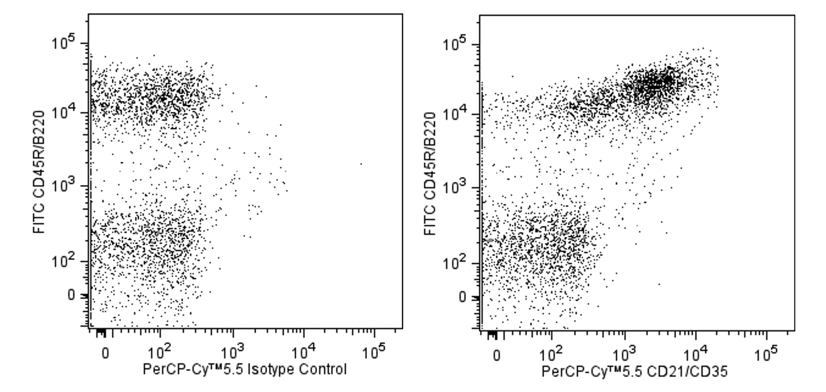
The 7G6 antibody recognizes an epitope shared by 145-150-kDa and 190-kDa complement receptor proteins, originally designated CR2 (CD21) and CR1 (CD35), respectively. In the mouse, CD21 and CD35 are expressed on the majority of peripheral B lymphocytes, on the majority of resident peritoneal macrophages and mast cells, on peripheral blood granulocytes after treatment with N-formyl-Met-Leu-Phe, and on follicular dendritic cells, but not on thymocytes, T cells, erythrocytes, or platelets. CD21 is a ligand-binding component of the CD19/CD21/CD81 signal-transduction complex associated with the antigen receptor on B lymphocytes. CD21/CD35 also co-localizes with CD19 on the surface of peritoneal mast cells. Cr2null mice display impaired inflammatory and humoral immune responses in vivo. The 7G6 mAb has been reported to inhibit rosette formation by C3d-bearing sheep erythrocytes, to block the complement dependent trapping of immune complexes by follicular dendritic cells, and to down-regulate mouse CD21/CD35 expression upon in vivo application, thus inhibiting primary antibody responses to immunization. Co-stimulation of B-cell differentiation via Sepharose-coupled 7G6 antibody has also been observed. The 7G6 mAb recognizes an epitope on CD35 distinct from the epitope recognized by anti-mouse CD35, clone 8C12, and it does not block binding of 8C12 mAb to mouse CD35.

Development References (19)
-
Ahearn JM, Fischer MB, Croix D, et al. Disruption of the Cr2 locus results in a reduction in B-1a cells and in an impaired B cell response to T-dependent antigen. Immunity. 1996; 4(3):251-262. (Biology). View Reference
-
Axcrona K, Gray D, Leanderson T. Regulation of B cell growth and differentiation via CD21 and CD40. Eur J Immunol. 1996; 26(9):2203-2207. (Biology). View Reference
-
Cariappa A, Tang M, Parng C, et al. The follicular versus marginal zone B lymphocyte cell fate decision is regulated by Aiolos, Btk, and CD21. Immunity. 2001; 14(5):603-615. (Biology). View Reference
-
Dempsey PW, Allison ME, Akkaraju S, Goodnow CC, Fearon DT. C3d of complement as a molecular adjuvant: bridging innate and acquired immunity. Science. 1996; 287(5247):348-350. (Biology). View Reference
-
Fagarasan S, Muramatsu M, Suzuki K, Nagaoka H, Hiai H, Honjo T. Critical roles of activation-induced cytidine deaminase in the homeostasis of gut flora. Science. 2002; 298(5597):1424-1427. (Clone-specific: Fluorescence microscopy). View Reference
-
Fischer MB, Goerg S, Shen L, et al. Dependence of germinal center B cells on expression of CD21/CD35 for survival. Science. 1998; 280(5363):582-585. (Biology). View Reference
-
Gommerman JL, Oh DY, Zhou X, et al. A role for CD21/CD35 and CD19 in responses to acute septic peritonitis: a potential mechanism for mast cell activation. J Immunol. 2000; 165(12):6915-6921. (Biology). View Reference
-
Heyman B, Wiersma EJ, Kinoshita T. In vivo inhibition of the antibody response by a complement receptor-specific monoclonal antibody. J Exp Med. 1990; 172(2):665-668. (Biology). View Reference
-
Hu H, Martin BK, Weis JJ, Weis JH. Expression of the murine CD21 gene is regulated by promoter and intronic sequences. J Immunol. 1997; 158(10):4758-4768. (Biology). View Reference
-
Kinoshita T, Takeda J, Hong K, Kozono H, Sakai H, Inoue K. Monoclonal antibodies to mouse complement receptor type 1 (CR1). Their use in a distribution study showing that mouse erythrocytes and platelets are CR1-negative. J Immunol. 1988; 140(9):3066-3072. (Immunogen). View Reference
-
Kinoshita T, Thyphronitis G, Tsokos GC, et al. Characterization of murine complement receptor type 2 and its immunological cross-reactivity with type 1 receptor. Int Immunol. 1990; 2(7):651-659. (Biology). View Reference
-
Kurtz CB, O'Toole E, Christensen SM, Weis JH. The murine complement receptor gene family. IV. Alternative splicing of Cr2 gene transcripts predicts two distinct gene products that share homologous domains with both human CR2 and CR1. J Immunol. 1990; 144(9):3581-3591. (Biology). View Reference
-
Molina H, Holers VM, Li B, et al. Markedly impaired humoral immune response in mice deficient in complement receptors 1 and 2. Proc Natl Acad Sci U S A. 1996; 93(8):3357-3361. (Biology). View Reference
-
Oliver AM, Martin F, Gartland GL, Carter RH, Kearney JF. Marginal zone B cells exhibit unique activation, proliferative and immunoglobulin secretory responses. Eur J Immunol. 1997; 27(9):2366-2374. (Biology). View Reference
-
Oliver AM, Martin F, Kearney JF. IgMhighCD21high lymphocytes enriched in the splenic marginal zone generate effector cells more rapidly than the bulk of follicular B cells. J Immunol. 1999; 162(12):7198-7207. (Biology). View Reference
-
Tedder TF, Zhou LJ, Engel P. The CD19/CD21 signal transduction complex of B lymphocytes. Immunol Today. 1994; 15(9):437-442. (Biology). View Reference
-
Thyphronitis G, Kinoshita T, Inoue K, et al. Modulation of mouse complement receptors 1 and 2 suppresses antibody responses in vivo. J Immunol. 1991; 147(1):224-230. (Biology). View Reference
-
Wiersma EJ, Kinoshita T, Heyman B. Inhibition of immunological memory and T-independent humoral responses by monoclonal antibodies specific for murine complement receptors. Eur J Immunol. 1991; 21(10):2501-2506. (Biology). View Reference
-
Yoshida K, van den Berg TK, Dijkstra CD. Two functionally different follicular dendritic cells in secondary lymphoid follicles of mouse spleen, as revealed by CR1/2 and FcR gamma II-mediated immune-complex trapping. Immunology. 1993; 80(1):34-39. (Biology). View Reference
Please refer to Support Documents for Quality Certificates
Global - Refer to manufacturer's instructions for use and related User Manuals and Technical data sheets before using this products as described
Comparisons, where applicable, are made against older BD Technology, manual methods or are general performance claims. Comparisons are not made against non-BD technologies, unless otherwise noted.
For Research Use Only. Not for use in diagnostic or therapeutic procedures.