Skip to main content Skip to navigation
V450 Mouse Anti-Bcl-6
Flow cytometric analysis of Bcl-6 expression in Ramos cells (Left Panel). Human Ramos cells were fixed with BD Cytofix™ Fixation Buffer (Cat. No. 554655) and permeabilized with BD Phosflow™ Perm Buffer III (Cat. No. 558050) followed by intracellular staining with either BD Horizon™ V450 Mouse Anti-Human Bcl-6 antibody (Cat. No. 562204, solid line histogram) or a BD Horizon™ V450 mIgG1, κ Isotype Control (Cat. No. 560373; dashed line histogram). Flow cytometric fluorescence histograms were derived from gated events with the forward and side light-scatter characteristics of viable cells. Flow cytometry was performed using a BD™ LSR II Flow Cytometer System.
V450 Mouse Anti-Bcl-6
Multicolor flow cytometric analysis of Bcl-6 expression in mouse B lymphocytes (Middle and Right Panel). BALB/c mouse mesenteric lymph node cells were stained with APC Rat Anti-Mouse B220 (Cat. No. 553092/561880), PerCP-Cy™5.5 Rat Anti-Mouse CD4 (Cat. No. 550954/561115), FITC Hamster Anti-Mouse Fas/CD95 (Cat. No. 554257), and PE Rat Anti-Mouse IgD (Cat. No. 558597) antibodies. Cells were washed, resuspended in RPMI with 10% FBS, and fixed with BD Phosflow™ Lyse/Fix Buffer (Cat. No. 558049). Cells were permeabilized with BD Phosflow™ Perm/Wash Buffer I (Cat. No. 557885), followed by intracellular staining with BD Horizon™ V450 Mouse Anti-Bcl-6 antibody (Cat. No. 562204). A two-color flow cytometric dot plot shows the expression of IgD versus Fas/CD95 by B cells identified as CD4-B220+ from gated events with the forward and side light-scatter characteristics of intact lymphocytes (Middle Panel). Germinal center (GC) B cells were identified as Fas/CD95-positive B lymphocytes that expressed low levels of IgD (IgDloCD95/Fas+) whereas non-GC B cells primarily expressed intermediate to high levels of IgD and little or no CD95/Fas. Flow cytometric fluorescence histograms (Right Panel) derived from gated cell subpopulations show intracellular Bcl-6 staining levels for mouse GC B cells (solid line histogram) and non-GC B cells (dashed line histogram). Flow cytometry was performed using a BD™ LSR II Flow Cytometer System.
Flow cytometric analysis of Bcl-6 expression in Ramos cells (Left Panel). Human Ramos cells were fixed with BD Cytofix™ Fixation Buffer (Cat. No. 554655) and permeabilized with BD Phosflow™ Perm Buffer III (Cat. No. 558050) followed by intracellular staining with either BD Horizon™ V450 Mouse Anti-Human Bcl-6 antibody (Cat. No. 562204, solid line histogram) or a BD Horizon™ V450 mIgG1, κ Isotype Control (Cat. No. 560373; dashed line histogram). Flow cytometric fluorescence histograms were derived from gated events with the forward and side light-scatter characteristics of viable cells. Flow cytometry was performed using a BD™ LSR II Flow Cytometer System.
Multicolor flow cytometric analysis of Bcl-6 expression in mouse B lymphocytes (Middle and Right Panel). BALB/c mouse mesenteric lymph node cells were stained with APC Rat Anti-Mouse B220 (Cat. No. 553092/561880), PerCP-Cy™5.5 Rat Anti-Mouse CD4 (Cat. No. 550954/561115), FITC Hamster Anti-Mouse Fas/CD95 (Cat. No. 554257), and PE Rat Anti-Mouse IgD (Cat. No. 558597) antibodies. Cells were washed, resuspended in RPMI with 10% FBS, and fixed with BD Phosflow™ Lyse/Fix Buffer (Cat. No. 558049). Cells were permeabilized with BD Phosflow™ Perm/Wash Buffer I (Cat. No. 557885), followed by intracellular staining with BD Horizon™ V450 Mouse Anti-Bcl-6 antibody (Cat. No. 562204). A two-color flow cytometric dot plot shows the expression of IgD versus Fas/CD95 by B cells identified as CD4-B220+ from gated events with the forward and side light-scatter characteristics of intact lymphocytes (Middle Panel). Germinal center (GC) B cells were identified as Fas/CD95-positive B lymphocytes that expressed low levels of IgD (IgDloCD95/Fas+) whereas non-GC B cells primarily expressed intermediate to high levels of IgD and little or no CD95/Fas. Flow cytometric fluorescence histograms (Right Panel) derived from gated cell subpopulations show intracellular Bcl-6 staining levels for mouse GC B cells (solid line histogram) and non-GC B cells (dashed line histogram). Flow cytometry was performed using a BD™ LSR II Flow Cytometer System.
Product Details
Down Arrow Up Arrow


BD Horizon™
BCL6; B-cell lymphoma 6 protein; LAZ3; Laz-3, ZBTB27, ZNF51
Human (QC Testing), Mouse (Tested in Development)
Mouse IgG1, κ
Human Bcl-6 Recombinant Protein
Intracellular staining (flow cytometry) (Routinely Tested)
5 µl
AB_10926203
Aqueous buffered solution containing protein stabilizer and ≤0.09% sodium azide.
RUO


Preparation And Storage

Store undiluted at 4°C and protected from prolonged exposure to light. Do not freeze. The monoclonal antibody was purified from tissue culture supernatant or ascites by affinity chromatography. The antibody was conjugated with BD Horizon™ V450 under optimum conditions, and unreacted BD Horizon™ V450 was removed.

Recommended Assay Procedures

We validate the quality of each batch of the K112-91 antibody conjugate by flow cytometry on human cell lines.  Investigators may use the same cell lines as controls for their staining procedure, namely Ramos (Positive; ATCC CRL-1596) and Jurkat (Negative; ATCC TIB-152) human cell lines actively growing in log phase (do not overgrow).  Cells are fixed with BD Cytofix™ Fixation Buffer (Cat. No. 554655; 10 minutes at 37°C), permeabilized with BD Phosflow™ Perm Buffer III (Cat. No. 558050; 30 minutes on ice), and washed using BD Pharmingen™ Stain Buffer (Cat. No. 554656), followed by intracellular staining with Mouse anti-Bcl-6 for 45 minutes at room temperature.

Product Notices

  1. Please refer to www.bdbiosciences.com/us/s/resources for technical protocols.
  2. Caution: Sodium azide yields highly toxic hydrazoic acid under acidic conditions. Dilute azide compounds in running water before discarding to avoid accumulation of potentially explosive deposits in plumbing.
  3. For fluorochrome spectra and suitable instrument settings, please refer to our Multicolor Flow Cytometry web page at www.bdbiosciences.com/colors.
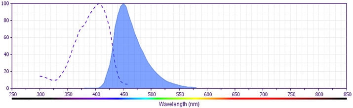
  4. BD Horizon V450 has a maximum absorption of 406 nm and maximum emission of 450 nm. Before staining with this reagent, please confirm that your flow cytometer is capable of exciting the fluorochrome and discriminating the resulting fluorescence.
  5. Pacific Blue™ is a trademark of Molecular Probes, Inc., Eugene, OR.
  6. This reagent has been pre-diluted for use at the recommended Volume per Test. We typically use 1 × 10^6 cells in a 100-µl experimental sample (a test).
  7. An isotype control should be used at the same concentration as the antibody of interest.
  8. This product is sold under license to the following patent: US Patent No. 6,174,997.
562204 Rev. 2
Antibody Details
Down Arrow Up Arrow
K112-91

The K112-91 monoclonal antibody specifically binds to Bcl-6. Bcl-6 was first identified as a proto-oncogene frequently deregulated by chromosomal translocations in non-Hodgkin B-cell lymphomas. It is a nuclear transcriptional repressor of the BTB/POZ zinc-finger family of transcription factors. In addition to its roles in cancer, Bcl-6 plays important roles in the differentiation of normal cells including B cells, thymocytes, CD4+ or CD8+ T cells. Bcl-6 is highly expressed in germinal center B cells, where it promotes the germinal center reaction by inducing proliferation and inhibiting the DNA-damage response. Bcl-6 has been identified as a key factor in promoting the differentiation of CD4+ follicular T helper (Tfh) cells that are involved in promoting germinal center formation and providing help to B cells. The interplay of Bcl-6 and another transcriptional repressor, Blimp-1, is thought to be critical in defining the results of both B-cell and T-cell differentiation.

The antibody is conjugated to BD Horizon™ V450, which has been developed for use in multicolor flow cytometry experiments and is available exclusively from BD Biosciences. It is excited by the Violet laser Ex max of 406 nm and has an Em Max at 450 nm. Conjugates with BD Horizon™ V450 can be used in place of Pacific Blue™ conjugates.

562204 Rev. 2
Format Details
Down Arrow Up Arrow
V450
BD Horizon™ V450 Dye is part of the BD Horizon™ violet family of dyes. This is a small organic fluorochrome with an excitation maximum (Ex Max) at 405-nm and an emission maximum (Em Max) at 450-nm. BD Horizon™ V450, driven by BD innovation, is designed to be excited by the violet laser (405 nm) and detected using an optical filter centered near 450-nm (e.g., a 450/50-nm bandpass filter). The dye can be excited by the UV (355-nm) laser resulting in cross-laser excitation and spillover. Please ensure that your instrument’s configurations (lasers and optical filters) are appropriate for this dye.
altImg
V450
Violet 405 nm
405 nm
450 nm
562204 Rev.2
Citations & References
Down Arrow Up Arrow

Development References (9)

  1. Baumjohann D, Okada T, Ansel KM. Cutting Edge: Distinct Waves of BCL6 Expression during T Follicular Helper Cell Development. J Immunol. 2011; 187(5):2089-2092. (Clone-specific: Flow cytometry). View Reference
  2. Crotty S, Choi YS, Kageyama R, et al. ICOS receptor instructs T follicular helper cell versus effector cell differentiation via induction of the transcriptional repressor Bcl6. Immunity. 2011; 34:1-15. (Clone-specific: Flow cytometry). View Reference
  3. Crotty S, Johnston RJ, Schoenberger SP. Effectors and memories: Bcl-6 and Blimp-1 in T and B lymphocyte differentiation. Nat Immunol. 2010; 11(2):114-120. (Biology). View Reference
  4. Crotty S. Follicular Helper CD4 T Cells (Tfh). Annu Rev Immunol. 2011; 29(1):621-663. (Biology). View Reference
  5. Fazilleau N, McHeyzer-Williams LJ, Rosen H, McHeyzer-Williams MG. The function of follicular helper T cells is regulated by the strength of T cell antigen receptor binding. Nat Rev Immunol. 2009; 10(4):375-384. (Biology). View Reference
  6. Johnston RJ, Poholek AC, DiToro D, et al. Bcl6 and Blimp-1 are reciprocal and antagonistic regulators of T follicular helper cell differentiation.. Science. 2009; 325(5943):1006-10. (Biology). View Reference
  7. Klein U, Dalla-Favera R. Germinal centres: role in B-cell physiology and malignancy. Nat Rev Immunol. 2008; 8(1):22-33. (Biology). View Reference
  8. Nurieva RI, Chung Y, Martinez GJ, et al. Bcl6 mediates the development of T follicular helper cells. Science. 2009; 325(5943):1001-1005. (Biology). View Reference
  9. Ye BH, Lista F, Lo Coco F, et al. Alterations of a zinc finger-encoding gene, BCL-6, in diffuse large-cell lymphoma. Science. 1993; 262(5134):747-750. (Biology). View Reference
View All (9) View Less
562204 Rev. 2

Please refer to Support Documents for Quality Certificates


Global - Refer to manufacturer's instructions for use and related User Manuals and Technical data sheets before using this products as described


Comparisons, where applicable, are made against older BD Technology, manual methods or are general performance claims.  Comparisons are not made against non-BD technologies, unless otherwise noted.

For Research Use Only. Not for use in diagnostic or therapeutic procedures.