

Preparation And Storage
Product Notices
- Since applications vary, each investigator should titrate the reagent to obtain optimal results.
- An isotype control should be used at the same concentration as the antibody of interest.
- Caution: Sodium azide yields highly toxic hydrazoic acid under acidic conditions. Dilute azide compounds in running water before discarding to avoid accumulation of potentially explosive deposits in plumbing.
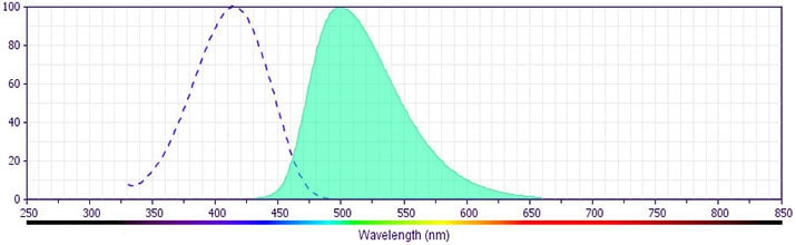
- BD Horizon V500 has a maximum absorption of 415 nm and maximum emission of 500 nm. Before staining with this reagent, please confirm that your flow cytometer is capable of exciting the fluorochrome and discriminating the resulting fluorescence.
- For fluorochrome spectra and suitable instrument settings, please refer to our Multicolor Flow Cytometry web page at www.bdbiosciences.com/colors.
- BD Horizon V500 is covered by one or more of the following US patents: 8,431,416.
- Species cross-reactivity detected in product development may not have been confirmed on every format and/or application.
- Please refer to www.bdbiosciences.com/us/s/resources for technical protocols.
Companion Products


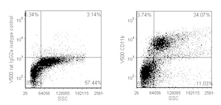
The M1/70 monoclonal antibody specifically binds to CD11b, also known as Integrin alpha M (Itgam or αM). CD11b is a 170-kDa type 1 transmembrane glycoprotein and belongs to the Integrin alpha chain family. CD11b serves as the alpha chain of the heterodimeric Mac-1 integrin (CD11b/CD18, αMβ2), also known as complement receptor 3 (CR3). Mac-1 mediates adhesion to ICAM-1 (CD54), ICAM-2 (CD102), fibrinogen and binding to C3bi. Mac-1 is expressed at varying levels on granulocytes, macrophages, myeloid-derived dendritic cells, natural killer cells, microglia, and B-1 B lymphocytes. Mac-1 expression is rapidly upregulated on neutrophils after activation, in the same time period that CD62L (L-selectin) is shed from the cell surface. The M1/70 antibody reportedly blocks cell adherence and C3bi binding but does not block cell-mediated lysis. Cross-reaction of the M1/70 antibody with CD11b expressed on human monocytes, polymorphonuclear leukocytes, and NK cells has been reported.
The antibody is conjugated to BD Horizon™ V500, which has been developed for use in multicolor flow cytometry experiments and is
available exclusively from BD Biosciences. It is excited by the Violet laser with an Ex max of 415 nm and Em Max at 500 nm. BD Horizon V500 conjugates emit at a similar wavelength to Amcyan yet exhibit reduced spillover into the FITC channel. For more information on BD Horizon V500, visit bdbiosciences.com/colors.
When compensating dyes in this spectral range (such as Horizon™ V500 and AmCyan), the most accurate compensation can be obtained using single stained cellular controls. Due to spectral differences between cells and beads in this channel, using BD CompBeads can result in spillover errors for V500 and AmCyan reagents. Therefore, the use of BD CompBeads or BD CompBeads Plus to determine spillover values for these reagents is not recommended. Different V500 reagents (e.g. CD4 vs. CD45) can have slightly different fluorescence spillover therefore, it may also be necessary to use clone specific compensation controls when using these reagents.
Development References (6)
-
Ault KA, Springer TA. Cross-reaction of a rat-anti-mouse phagocyte-specific monoclonal antibody (anti-Mac-1) with human monocytes and natural killer cells. J Immunol. 1981; 126(1):359-364. (Immunogen: Immunoprecipitation). View Reference
-
Greimers R, Trebak M, Moutschen M, Jacobs N, Boniver J. Improved four-color flow cytometry method using fluo-3 and triple immunofluorescence for analysis of intracellular calcium ion ([Ca2+]i) fluxes among mouse lymph node B- and T-lymphocyte subsets. Cytometry. 1996; 23(3):205-217. (Methodology: Flow cytometry). View Reference
-
Lagasse E, Weissman IL. Flow cytometric identification of murine neutrophils and monocytes. J Immunol Methods. 1996; 197(1-2):139-150. (Methodology: Flow cytometry). View Reference
-
Springer T, Galfre G, Secher DS, Milstein C. Mac-1: a macrophage differentiation antigen identified by monoclonal antibody. Eur J Immunol. 1979; 9(4):301-306. (Clone-specific: Immunoprecipitation). View Reference
-
Springer T, Galfre G, Secher DS, Milstein C. Monoclonal xenogeneic antibodies to murine cell surface antigens: identification of novel leukocyte differentiation antigens. Eur J Immunol. 1978; 8(8):539-551. (Immunogen: Immunoprecipitation). View Reference
-
Springer TA, Davignon D, Ho MK, Kurzinger K, Martz E, Sanchez-Madrid F. LFA-1 and Lyt-2,3, molecules associated with T lymphocyte-mediated killing; and Mac-1, an LFA-1 homologue associated with complement receptor function. Immunol Rev. 1982; 68:171-195. (Biology). View Reference
Please refer to Support Documents for Quality Certificates
Global - Refer to manufacturer's instructions for use and related User Manuals and Technical data sheets before using this products as described
Comparisons, where applicable, are made against older BD Technology, manual methods or are general performance claims. Comparisons are not made against non-BD technologies, unless otherwise noted.
For Research Use Only. Not for use in diagnostic or therapeutic procedures.