
Preparation And Storage
Product Notices
- Since applications vary, each investigator should titrate the reagent to obtain optimal results.
- Please refer to www.bdbiosciences.com/us/s/resources for technical protocols.
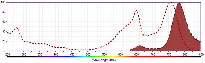
- APC-Cy7 is a tandem fluorochrome composed of Allophycocyanin (APC), which is excited by laser lines between 595 and 647 nm and serves as an energy donor, coupled to the cyanine dye Cy7™, which acts as an energy acceptor and fluoresces at 780 nm. BD Biosciences Pharmingen has maximized the fluorochrome energy transfer in APC-Cy7, thus maximizing its fluorescence emission intensity, minimizing residual emission from APC, and minimizing required electronic compensation in multilaser-laser flow cytometry systems. Note: Although every effort is made to minimize the lot-to-lot variation in residual emission from APC, it is strongly recommended that every lot be tested for differences in the amount of compensation required and that individual compensation controls are run for each APC-Cy7 conjugate.
- APC-Cy7 tandem fluorochrome emission is collected in a detector for fluorescence wavelengths of 750 nm and higher.
- Please observe the following precautions: Absorption of visible light can significantly alter the energy transfer occurring in any tandem fluorochrome conjugate; therefore, we recommend that special precautions be taken (such as wrapping vials, tubes, or racks in aluminum foil) to prevent exposure of conjugated reagents, including cells stained with those reagents, to room illumination.
- Cy is a trademark of GE Healthcare.
- Warning: Some APC-Cy7 and PE-Cy7 conjugates show changes in their emission spectrum with prolonged exposure to formaldehyde. If you are unable to analyze fixed samples within four hours, we recommend that you use BD™ Stabilizing Fixative (Cat. No. 338036).
- Caution: Sodium azide yields highly toxic hydrazoic acid under acidic conditions. Dilute azide compounds in running water before discarding to avoid accumulation of potentially explosive deposits in plumbing.
- For fluorochrome spectra and suitable instrument settings, please refer to our Multicolor Flow Cytometry web page at www.bdbiosciences.com/colors.
- An isotype control should be used at the same concentration as the antibody of interest.
Companion Products


The 53-2.1 monoclonal antibody specifically binds to the CD90.2 (Thy-1.2) alloantigen on thymocytes, most peripheral T lymphocytes, some intraepithelial T lymphocytes (IEL, DEC), epithelial cells, fibroblasts, neurons, hematopoietic stem cells, but not B lymphocytes, of most mouse strains. The 53-2.1 antibody has been reported not to crossreact with Thy-1.1 (e.g., AKR/J, PL), or with rat Thy-1. CD90 is a glycophosphatidylinositol-anchored membrane glycoprotein of the Ig superfamily that is involved in signal transduction. In addition, there is evidence that CD90 mediates adhesion of thymocytes to thymic stroma. The 53-2.1 antibody has been reported to block the binding of the Rat Anti-Mouse CD90.2 antibody (Clone 30-H12) to immobilized thymocyte membranes.
Development References (13)
-
Borrello MA, Phipps RP. Differential Thy-1 expression by splenic fibroblasts defines functionally distinct subsets. Cell Immunol. 1996; 173(2):198-206. (Biology). View Reference
-
He HT, Naquet P, Caillol D, Pierres M. Thy-1 supports adhesion of mouse thymocytes to thymic epithelial cells through a Ca2(+)-independent mechanism. J Exp Med. 1991; 173(2):515-518. (Biology). View Reference
-
Hueber AO, Raposo G, Pierres M, He HT. Thy-1 triggers mouse thymocyte apoptosis through a bcl-2-resistant mechanism. J Exp Med. 1994; 179(3):785-796. (Biology). View Reference
-
Ikuta K, Uchida N, Friedman J, Weissman IL. Lymphocyte development from stem cells. Annu Rev Immunol. 1992; 10:759-783. (Biology). View Reference
-
Kroczek RA, Gunter KC, Germain RN, Shevach EM. Thy-1 functions as a signal transduction molecule in T lymphocytes and transfected B lymphocytes. Nature. 1986; 322(6075):181-184. (Biology). View Reference
-
LeFrancois L. Extrathymic differentiation of intraepithelial lymphocytes: generation of a separate and unequal T-cell repertoire. Immunol Today. 1991; 12(12):436-438. (Biology). View Reference
-
Ledbetter JA, Herzenberg LA. Xenogeneic monoclonal antibodies to mouse lymphoid differentiation antigens. Immunol Rev. 1979; 47:63-90. (Immunogen: Blocking). View Reference
-
Ledbetter JA, Rouse RV, Micklem HS, Herzenberg LA. T cell subsets defined by expression of Lyt-1,2,3 and Thy-1 antigens. Two-parameter immunofluorescence and cytotoxicity analysis with monoclonal antibodies modifies current views. J Exp Med. 1980; 152(2):280-295. (Biology). View Reference
-
Radrizzani M, Carminatti H, Pivetta OH, Idoyaga Vargas VP. Developmental regulation of Thy 1.2 rate of synthesis in the mouse cerebellum. J Neurosci Res. 1995; 42(2):220-227. (Biology). View Reference
-
Tigelaar RE, Lewis JM, Bergstresser PR. TCR gamma/delta+ dendritic epidermal T cells as constituents of skin-associated lymphoid tissue. J Invest Dermatol. 1990; 94(6):58S-63S. (Biology). View Reference
-
Williams AF, Gagnon J. Neuronal cell Thy-1 glycoprotein: homology with immunoglobulin. Science. 1982; 216(4547):696-703. (Biology). View Reference
-
Zheng B, Han S, Kelsoe G. T helper cells in murine germinal centers are antigen-specific emigrants that downregulate Thy-1. J Exp Med. 1996; 184(3):1083-1091. (Biology). View Reference
-
Zhong RK, Donnenberg AD, Edison L, Harrison DE. The appearance of Thy-1- donor T cells in the peripheral circulation 3-6 weeks after bone marrow transplantation suggests an extrathymic origin. Int Immunol. 1996; 8(2):171-176. (Biology). View Reference
Please refer to Support Documents for Quality Certificates
Global - Refer to manufacturer's instructions for use and related User Manuals and Technical data sheets before using this products as described
Comparisons, where applicable, are made against older BD Technology, manual methods or are general performance claims. Comparisons are not made against non-BD technologies, unless otherwise noted.
For Research Use Only. Not for use in diagnostic or therapeutic procedures.