-
Reagents
- Flow Cytometry Reagents
-
Western Blotting and Molecular Reagents
- Immunoassay Reagents
-
Single-Cell Multiomics Reagents
- BD® AbSeq Assay
- BD Rhapsody™ Accessory Kits
- BD® Single-Cell Multiplexing Kit
- BD Rhapsody™ Targeted mRNA Kits
- BD Rhapsody™ Whole Transcriptome Analysis (WTA) Amplification Kit
- BD Rhapsody™ TCR/BCR Profiling Assays for Human and Mouse
- BD® OMICS-Guard Sample Preservation Buffer
- BD Rhapsody™ ATAC-Seq Assays
-
Functional Assays
-
Microscopy and Imaging Reagents
-
Cell Preparation and Separation Reagents
-
Training
- Flow Cytometry Basic Training
-
Product-Based Training
- BD FACSDiscover™ S8 Cell Sorter Product Training
- Accuri C6 Plus Product-Based Training
- FACSAria Product Based Training
- FACSCanto Product-Based Training
- FACSLyric Product-Based Training
- FACSMelody Product-Based Training
- FACSymphony Product-Based Training
- HTS Product-Based Training
- LSRFortessa Product-Based Training
- Advanced Training
-
- BD® AbSeq Assay
- BD Rhapsody™ Accessory Kits
- BD® Single-Cell Multiplexing Kit
- BD Rhapsody™ Targeted mRNA Kits
- BD Rhapsody™ Whole Transcriptome Analysis (WTA) Amplification Kit
- BD Rhapsody™ TCR/BCR Profiling Assays for Human and Mouse
- BD® OMICS-Guard Sample Preservation Buffer
- BD Rhapsody™ ATAC-Seq Assays
-
- BD FACSDiscover™ S8 Cell Sorter Product Training
- Accuri C6 Plus Product-Based Training
- FACSAria Product Based Training
- FACSCanto Product-Based Training
- FACSLyric Product-Based Training
- FACSMelody Product-Based Training
- FACSymphony Product-Based Training
- HTS Product-Based Training
- LSRFortessa Product-Based Training
- United States (English)
-
Change country/language
Old Browser
This page has been recently translated and is available in French now.
Looks like you're visiting us from {countryName}.
Would you like to stay on the current country site or be switched to your country?


Flow cytometric analysis of S6 (pS235/pS236) expression in activated human peripheral blood lymphocytes. Human peripheral blood mononuclear cells were either left untreated (dashed line histogram) or treated with Phorbol 12-Myristate 13-Acetate (PMA; Sigma-Aldrich, Cat. No. P-8139) at 50 nM for 30 minutes (solid line histogram). The cells were then fixed in BD Cytofix™ Fixation Buffer (Cat. No. 554655) at 37°C for 10 minutes, permeabilized with BD Phosflow™ Perm Buffer III (Cat. No. 558050) on ice for 30 minutes, and stained with BD Horizon™ V450 Mouse anti-S6 (pS235/pS236) antibody (Cat. No. 561457). The fluorescence histograms were derived from events with the forward and side light-scatter characteristics of intact lymphocytes. Flow cytometry was performed using a BD™ LSR II Flow Cytometer System.


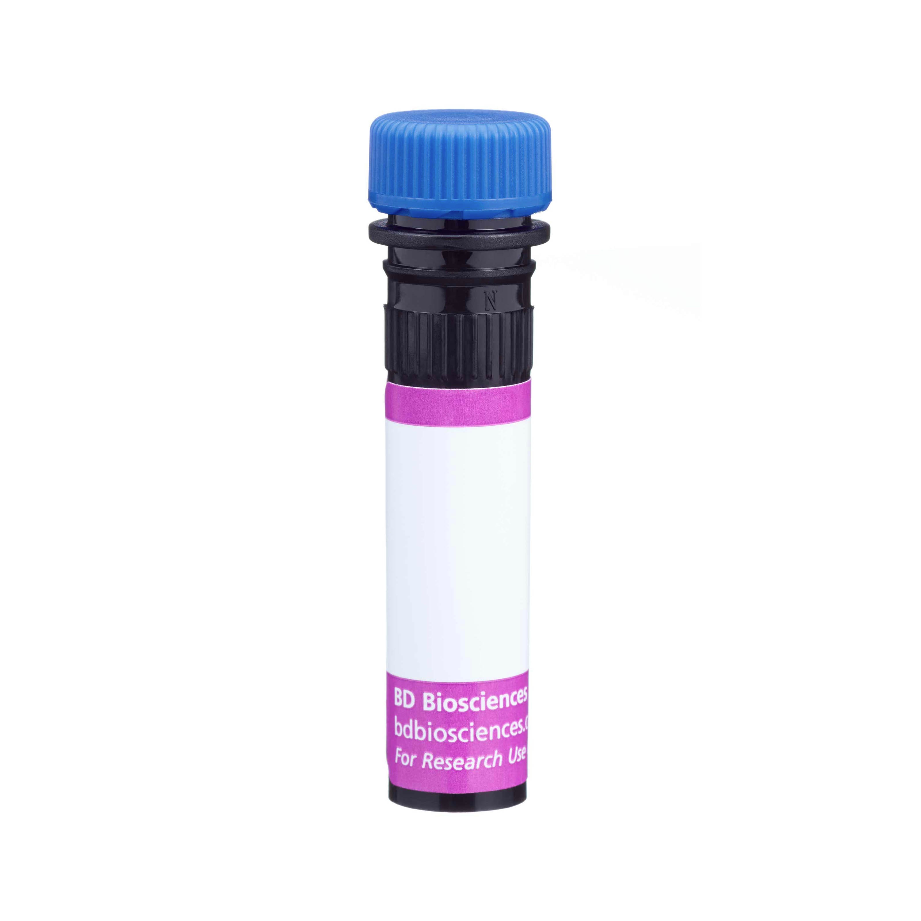
BD™ Phosflow V450 Mouse anti-S6 (pS235/pS236)

Regulatory Status Legend
Any use of products other than the permitted use without the express written authorization of Becton, Dickinson and Company is strictly prohibited.
Preparation And Storage
Product Notices
- This reagent has been pre-diluted for use at the recommended Volume per Test. We typically use 1 × 10^6 cells in a 100-µl experimental sample (a test).
- Caution: Sodium azide yields highly toxic hydrazoic acid under acidic conditions. Dilute azide compounds in running water before discarding to avoid accumulation of potentially explosive deposits in plumbing.
- BD Horizon V450 has a maximum absorption of 406 nm and maximum emission of 450 nm. Before staining with this reagent, please confirm that your flow cytometer is capable of exciting the fluorochrome and discriminating the resulting fluorescence.
- Pacific Blue™ is a trademark of Molecular Probes, Inc., Eugene, OR.
- For fluorochrome spectra and suitable instrument settings, please refer to our Multicolor Flow Cytometry web page at www.bdbiosciences.com/colors.
- Please refer to www.bdbiosciences.com/us/s/resources for technical protocols.
Companion Products


Ribosomal protein S6 (~29 kDa calculated and ~32 kDa observed molecular weights) is a component of the 40S ribosomal subunit and belongs to the S6E family of ribosomal proteins. The S6 ribosomal protein plays a role in regulating the translation of RNAs and thus controlling the growth and proliferation of cells. S6 ribosomal protein phosphorylation, especially at multiple C-terminal serine residues S235, S236, S240, and S244, activates S6. The activated S6 ribosomal protein in turn upregulates the ribosomal translation of RNA species coding for other ribosomal proteins, peptide elongation factors and other proteins involved in cell cycle entry and progression. These phosphorylations are mediated by various kinases (e.g., p70S6K and PKCD) activated through cellular responses to growth factors, cytokines, tumor promoting agents, and mitogens. The S6 ribosomal protein can be dephosphorylated in growth-arrested cells.
The N7-548 monoclonal antibody specifically detects the S6 ribosomal protein phosphorylated at S235 and S236.
The antibody is conjugated to BD Horizon™ V450, which has been developed for use in multicolor flow cytometry experiments and is available exclusively from BD Biosciences. It is excited by the Violet laser Ex max of 406 nm and has an Em Max at 450 nm. Conjugates with BD Horizon™ V450 can be used in place of Pacific Blue™ conjugates.

Development References (9)
-
Blatt K, Herrmann H, Mirkina I, et al. The PI3-kinase/mTOR-targeting drug NVP-BEZ235 inhibits growth and IgE-dependent activation of human mast cells and basophils. PLoS ONE. 2012; 7(1):e29925. (Clone-specific: Flow cytometry). View Reference
-
Corradetti MN, Inoki K, Guan KL. The stress-inducted proteins RTP801 and RTP801L are negative regulators of the mammalian target of rapamycin pathway. J Biol Chem. 2005; 280(11):9769-9772. (Biology). View Reference
-
Gu JJ, Santiago L, Mitchell BS. Synergy between imatinib and mycophenolic acid in inducing apoptosis in cell lines expressing Bcr-Abl. Blood. 2005; 105(8):3270-3277. (Biology). View Reference
-
Jastrzebski K, Hannan KM, Tchoubrieva EB, Hannan RD, Pearson RB. Coordinate regulation of ribosome biogenesis and function by the ribosomal protein S6 kinase, a key mediator of mTOR function. Growth Factors. 2007; 25(4):209-226. (Biology). View Reference
-
Kleijn M, Proud CG. The regulation of protein synthesis and translation factors by CD3 and CD28 in human primary T lymphocytes. BMC Biochem. 2002; 3:11. (Biology). View Reference
-
Lal L, Li Y, Smith J, et al. Activation of the p70 S6 kinase by all-trans-retinoic acid in acute promyelocytic leukemia cells.. Blood. 2005; 105(4):1669-77. (Biology). View Reference
-
Shah OJ, Ghosh S, Hunter T. Mitotic regulation of ribosomal S6 kinase 1 involves Ser/Thr, Pro phosphorylation of consensus and non-consensus sites by Cdc2. J Biol Chem. 2003; 278(18):16433-16442. (Biology). View Reference
-
Shah OJ, Hunter T. Critical role of T-loop and H-motif phosphorylation in the regulation of S6 kinase 1 by the tuberous sclerosis complex. J Biol Chem. 2004; 279(20):20816-20823. (Biology). View Reference
-
van de Laar L, van den Bosch A, Boonstra A, et al. PI3K-PKB hyperactivation augments human plasmacytoid dendritic cell development and function. Blood. 2012; 120(25):4982-4991. (Clone-specific: Flow cytometry). View Reference
Please refer to Support Documents for Quality Certificates
Global - Refer to manufacturer's instructions for use and related User Manuals and Technical data sheets before using this products as described
Comparisons, where applicable, are made against older BD Technology, manual methods or are general performance claims. Comparisons are not made against non-BD technologies, unless otherwise noted.
For Research Use Only. Not for use in diagnostic or therapeutic procedures.
Report a Site Issue
This form is intended to help us improve our website experience. For other support, please visit our Contact Us page.