
Preparation And Storage
Recommended Assay Procedures
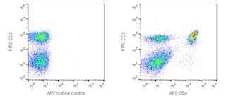
Flow cytometry: The JES6-5H4 antibody is useful for immunofluorescent staining and flow cytometric analysis to identify and enumerate IL-2 producing cells within mixed cell populations. A useful control investigators may consider using for demonstrating specificity of staining, is to pre-block with one of the following reagents: (1) recombinant mouse IL-2 (Cat. No. 550069) or (2) unlabeled JES6-5H4 antibody (Cat. No. 554425), prior to staining.
Product Notices
- Since applications vary, each investigator should titrate the reagent to obtain optimal results.
- An isotype control should be used at the same concentration as the antibody of interest.
- Please refer to www.bdbiosciences.com/us/s/resources for technical protocols.
- The Alexa Fluor®, Pacific Blue™, and Cascade Blue® dye antibody conjugates in this product are sold under license from Molecular Probes, Inc. for research use only, excluding use in combination with microarrays, or as analyte specific reagents. The Alexa Fluor® dyes (except for Alexa Fluor® 430), Pacific Blue™ dye, and Cascade Blue® dye are covered by pending and issued patents.
- Alexa Fluor® 700 has an adsorption maximum of ~700nm and a peak fluorescence emission of ~720nm. Before staining cells with this reagent, please confirm that your flow cytometer is capable of exciting the fluorochrome and discriminating the resulting fluorescence.
- Alexa Fluor® is a registered trademark of Molecular Probes, Inc., Eugene, OR.
- Caution: Sodium azide yields highly toxic hydrazoic acid under acidic conditions. Dilute azide compounds in running water before discarding to avoid accumulation of potentially explosive deposits in plumbing.
- For fluorochrome spectra and suitable instrument settings, please refer to our Multicolor Flow Cytometry web page at www.bdbiosciences.com/colors.
Companion Products


.png?imwidth=320)
The JES6-5H4 monoclonal antibody specifically binds to mouse interleukin-2 (IL-2), a multifunctional cytokine that plays pivotal roles in immunity and tolerance. It is produced by activated T cells and affects the activation, growth, proliferation and/or differentiation of various cell types including T and B lymphocytes and their precursors, LAK cells, NK cells, and monocytes/macrophages. IL-2 mediates its biological activities by binding to IL-2 receptor complexes. The intermediate affinity IL-2R is comprised of IL-2Rβ (CD122) and common gamma chain (γc; CD132) subunits, whereas the high-affinity IL-2R is comprised of IL-2Rα (CD25), IL-2Rβ, and γc subunits. The JES6-5H4 monoclonal antibody binds to IL-2 and neutralizes its biological activity.
Development References (12)
-
Awatsuji H, Furukawa Y, Nakajima M, Furukawa S, Hayashi K.. Interleukin-2 as a neurotrophic factor for supporting the survival of neurons cultured from various regions of fetal rat brain. J Neurosci Res. 1993; 35(3):305-311. (Biology). View Reference
-
Gillis S, Ferm MM, Ou W, Smith KA. T cell growth factor: parameters of production and a quantitative microassay for activity. J Immunol. 1978; 120(6):2027-2032. (Biology). View Reference
-
Gillis S, Ferm MM, Ou W, Smith KA. T cell growth factor: parameters of production and a quantitative microassay for activity. J Immunol. 1978; 120(6):2027-2032. (Biology). View Reference
-
Helms T, Boehm BO, Asaad RJ, Trezza RP, Lehmann PV, Tary-Lehmann M. Direct visualization of cytokine-producing recall antigen-specific CD4 memory T cells in healthy individuals and HIV patients. J Immunol. 2000; 164(7):3723-3732. (Biology). View Reference
-
Kashima N, Nishi-Takaoka C, Fujita T, et al. Unique structure of murine interleukin-2 as deduced from cloned cDNAs. Nature. 1985; 313(6001):402-404. (Biology). View Reference
-
Kubo M, Cinader B. Polymorphism of age-related changes in interleukin (IL) production: differential changes of T helper subpopulations, synthesizing IL 2, IL 3 and IL 4. Eur J Immunol. 1990; 20(6):1289-1296. (Biology). View Reference
-
Mochizuki DY, Watson J, Gillis S. Biochemical separation of interleukin 2. J Immunol Methods. 1980; 39(3):185-201. (Biology). View Reference
-
Mosmann TR, Cherwinski H, Bond MW, Giedlin MA, Coffman RL. Two types of murine helper T cell clone. I. Definition according to profiles of lymphokine activities and secreted proteins. J Immunol. 1986; 136(7):2348-2357. (Biology). View Reference
-
Prussin C, Metcalfe DD. Detection of intracytoplasmic cytokine using flow cytometry and directly conjugated anti-cytokine antibodies. J Immunol Methods. 1995; 188(1):117-128. (Methodology: Flow cytometry). View Reference
-
Sander B, Hoiden I, Andersson U, Moller E, Abrams JS. Similar frequencies and kinetics of cytokine producing cells in murine peripheral blood and spleen. Cytokine detection by immunoassay and intracellular immunostaining. J Immunol Methods. 1993; 166(2):201-214. (Methodology: ELISA). View Reference
-
Watson J, Mochizuki D. Interleukin 2: a class of T cell growth factors. Immunol Rev. 1980; 51:257-278. (Biology). View Reference
-
Yokota T, Arai N, Lee F, Rennick D, Mosmann T, Arai K. Use of a cDNA expression vector for isolation of mouse interleukin 2 cDNA clones: expression of T-cell growth-factor activity after transfection of monkey cells. Proc Natl Acad Sci U S A. 1985; 82(1):68-72. (Biology). View Reference
Please refer to Support Documents for Quality Certificates
Global - Refer to manufacturer's instructions for use and related User Manuals and Technical data sheets before using this products as described
Comparisons, where applicable, are made against older BD Technology, manual methods or are general performance claims. Comparisons are not made against non-BD technologies, unless otherwise noted.
For Research Use Only. Not for use in diagnostic or therapeutic procedures.