
Preparation And Storage
Recommended Assay Procedures
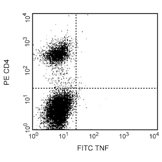
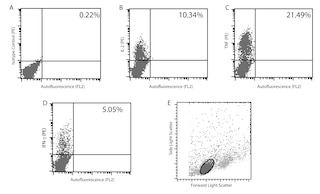
Flow cytometry: The MP6-XT22 antibody is useful for immunofluorescent staining and flow cytometric analysis to identify and enumerate TNF producing cells within mixed cell populations. A useful control investigators may consider using for demonstrating specificity of staining, is to pre-block with one of the following reagents: (1) recombinant mouse TNF (Cat. No. 554589) or (2) unlabeled MP6-XT22 antibody (Cat. No. 554416), prior to staining.
Cell Preparation: Investigators not wishing to utilize MiCK-1 cells may alternatively prepare mouse splenocytes (e.g BALB/c) stimulated for 4-6 hours with PMA (5 ng/mL, Sigma-Aldrich Cat. No. P-8139) and ionomycin (500 ng, Sigma-Aldrich Cat. No. I-0634) in the presence of 1 µg/mL Protein Transport Inhibitor [containing Brefeldin A] (Cat. No. 555029). Investigators are advised to fix and permeabilize the cells with BD Cytofix/CytoPerm Plus Kit (Cat. No. 555028) prior to staining.
Product Notices
- Since applications vary, each investigator should titrate the reagent to obtain optimal results.
- An isotype control should be used at the same concentration as the antibody of interest.
- Caution: Sodium azide yields highly toxic hydrazoic acid under acidic conditions. Dilute azide compounds in running water before discarding to avoid accumulation of potentially explosive deposits in plumbing.
- Warning: Some APC-Cy7 and PE-Cy7 conjugates show changes in their emission spectrum with prolonged exposure to formaldehyde. If you are unable to analyze fixed samples within four hours, we recommend that you use BD™ Stabilizing Fixative (Cat. No. 338036).
- Please observe the following precautions: Absorption of visible light can significantly alter the energy transfer occurring in any tandem fluorochrome conjugate; therefore, we recommend that special precautions be taken (such as wrapping vials, tubes, or racks in aluminum foil) to prevent exposure of conjugated reagents, including cells stained with those reagents, to room illumination.
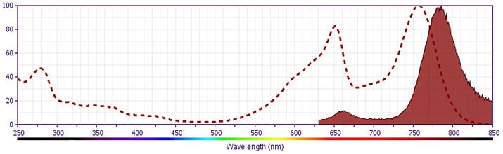
- APC-Cy7 tandem fluorochrome emission is collected in a detector for fluorescence wavelengths of 750 nm and higher.
- APC-Cy7 is a tandem fluorochrome composed of Allophycocyanin (APC), which is excited by laser lines between 595 and 647 nm and serves as an energy donor, coupled to the cyanine dye Cy7™, which acts as an energy acceptor and fluoresces at 780 nm. BD Biosciences Pharmingen has maximized the fluorochrome energy transfer in APC-Cy7, thus maximizing its fluorescence emission intensity, minimizing residual emission from APC, and minimizing required electronic compensation in multilaser-laser flow cytometry systems. Note: Although every effort is made to minimize the lot-to-lot variation in residual emission from APC, it is strongly recommended that every lot be tested for differences in the amount of compensation required and that individual compensation controls are run for each APC-Cy7 conjugate.
- For fluorochrome spectra and suitable instrument settings, please refer to our Multicolor Flow Cytometry web page at www.bdbiosciences.com/colors.
- Cy is a trademark of GE Healthcare.
- Please refer to www.bdbiosciences.com/us/s/resources for technical protocols.
Companion Products


The MP6-XT22 antibody specifically binds to mouse Tumor Necrosis Factor (TNF, also known as TNF-α). TNF is produced by many activated cell types including monocytes, macrophages, astrocytes, granulocytes, mast cells, T and B lymphocytes, NK cells, keratinocytes, fibroblasts, adipocytes, and certain tumor cells. Activated cells express type II transmembrane TNF glycoproteins that associate as homotrimeric complexes. After enzymatic cleavage, the extracellular regions of membrane TNF are shed as soluble homotrimers. TNF is a potent multifunctional cytokine that can exert regulatory and cytotoxic effects on a wide range of normal lymphoid and non-lymphoid cells and tumor cells. Although TNF serves as a primary mediator in protective immune responses against microbial and viral pathogens, it can also drive systemic pathophysiologic responses including septic shock, cachexia and autoimmune diseases. Mouse TNF exerts its biological activities by binding and signaling through cell surface membrane Type I and Type II TNF Receptors (aka, TNFRI/CD120a and TNFRII/CD120b, respectively).
Development References (12)
-
Abrams JS, Roncarolo MG, Yssel H, Andersson U, Gleich GJ, Silver JE. Strategies of anti-cytokine monoclonal antibody development: immunoassay of IL-10 and IL-5 in clinical samples. Immunol Rev. 1992; 127:5-24. (Biology). View Reference
-
Drew PD, Chavis JA. Female sex steroids: effects upon microglial cell activation. J Neuroimmunol. 2000; 111(1-2):77-85. (Biology). View Reference
-
Drew PD, Chavis JA. Inhibition of microglial cell activation by cortisol. Brain Res Bull. 2000; 52(5):391-396. (Biology). View Reference
-
Drew PD, Chavis JA.. The cyclopentone prostaglandin 15-deoxy-Delta(12,14) prostaglandin J2 represses nitric oxide, TNF-alpha, and IL-12 production by microglial cells.. J Neuroimmunol. 2001; 115(1-2):28-35. (Biology). View Reference
-
Ferran C, Dautry F, Merite S, et al. Anti-tumor necrosis factor modulates anti-CD3-triggered T cell cytokine gene expression in vivo. J Clin Invest. 1994; 93(5):2189-2196. (Biology). View Reference
-
He J, Gurunathan S, Iwasaki A, Ash-Shaheed B, Kelsall BL. Primary role for Gi protein signaling in the regulation of interleukin 12 production and the induction of T helper cell type 1 responses . J Exp Med. 2000; 191(9):1605-1610. (Biology). View Reference
-
Leiby DA, Fortier AH, Crawford RM, Schreiber RD, Nacy CA. In vivo modulation of the murine immune response to Francisella tularensis LVS by administration of anticytokine antibodies. Infect Immun. 1992; 60(1):84-89. (Biology). View Reference
-
Merrick BA, He CY, Craig WA, et al. Two dimensional gel electrophoresis of cellular and secreted proteins from rat alveolar macrophages after lipopolysaccharide treatment. Appl Theor Electrophor. 1992; 2(6):177-187. (Biology). View Reference
-
Prussin C, Metcalfe DD. Detection of intracytoplasmic cytokine using flow cytometry and directly conjugated anti-cytokine antibodies. J Immunol Methods. 1995; 188(1):117-128. (Methodology: Flow cytometry). View Reference
-
Rabinovici R, Bugelski PJ, Esser KM, et al. Tumor necrosis factor-alpha mediates endotoxin-induced lung injury in platelet activating factor-primed rats. J Pharmacol Exp Ther. 1993; 267(3):1550-1557. (Biology). View Reference
-
Sheehan KC, Ruddle NH, Schreiber RD. Generation and characterization of hamster monoclonal antibodies that neutralize murine tumor necrosis factors. J Immunol. 1989; 142(11):3884-3893. (Biology). View Reference
-
Takahashi S, Kapas L, Fang J, Krueger JM. An anti-tumor necrosis factor antibody suppresses sleep in rats and rabbits. Brain Res. 1995; 690(2):241-244. (Biology). View Reference
Please refer to Support Documents for Quality Certificates
Global - Refer to manufacturer's instructions for use and related User Manuals and Technical data sheets before using this products as described
Comparisons, where applicable, are made against older BD Technology, manual methods or are general performance claims. Comparisons are not made against non-BD technologies, unless otherwise noted.
For Research Use Only. Not for use in diagnostic or therapeutic procedures.