-
Training
- Flow Cytometry Basic Training
-
Product-Based Training
- BD FACSDiscover™ S8 Cell Sorter Product Training
- Accuri C6 Plus Product-Based Training
- FACSAria Product Based Training
- FACSCanto Product-Based Training
- FACSLyric Product-Based Training
- FACSMelody Product-Based Training
- FACSymphony Product-Based Training
- HTS Product-Based Training
- LSRFortessa Product-Based Training
- Advanced Training
-
- BD FACSDiscover™ S8 Cell Sorter Product Training
- Accuri C6 Plus Product-Based Training
- FACSAria Product Based Training
- FACSCanto Product-Based Training
- FACSLyric Product-Based Training
- FACSMelody Product-Based Training
- FACSymphony Product-Based Training
- HTS Product-Based Training
- LSRFortessa Product-Based Training
- United States (English)
-
Change country/language
Old Browser
This page has been recently translated and is available in French now.
Looks like you're visiting us from {countryName}.
Would you like to stay on the current country site or be switched to your country?


Flow cytometric analysis of apoptotic and non-apoptotic populations for Active Caspase-3. Jurkat cells (Human T-cell leukemia; ATCC TIB-152) were left untreated (shaded) or were treated with 4-12 µM of camptothecin (Sigma-Aldrich Cat. No. C9911) for 4-6 hr to induce apoptosis (unshaded). Cells were washed once in 1X PBS, then fixed and permeabilized using the BD Cytofix/Cytoperm™ Kit (Cat. No. 554714) followed by staining with the Alexa Fluor® 647 Rabbit Anti-Active Caspase-3 antibody. Histograms were derived from gated events based on light scattering characteristics for Jurkat cells. Flow cytometry was performed on a BD™ LSR II flow cytometry system.


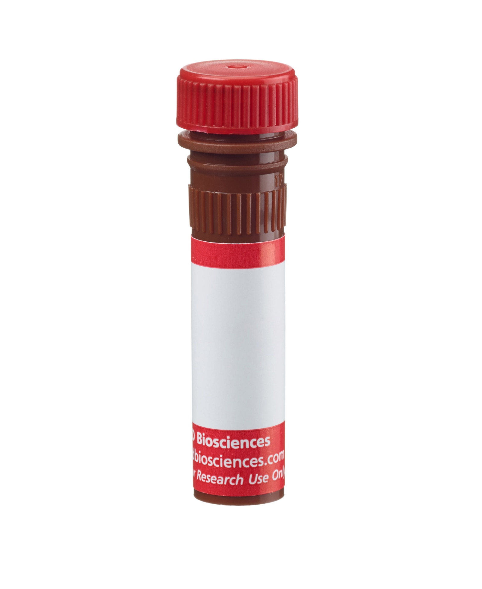
BD Pharmingen™ Alexa Fluor® 647 Rabbit Anti-Active Caspase-3

Regulatory Status Legend
Any use of products other than the permitted use without the express written authorization of Becton, Dickinson and Company is strictly prohibited.
Preparation And Storage
Product Notices
- This reagent has been pre-diluted for use at the recommended Volume per Test. We typically use 1 × 10^6 cells in a 100-µl experimental sample (a test).
- An isotype control should be used at the same concentration as the antibody of interest.
- Alexa Fluor® 647 fluorochrome emission is collected at the same instrument settings as for allophycocyanin (APC).
- Alexa Fluor® is a registered trademark of Molecular Probes, Inc., Eugene, OR.
- The Alexa Fluor®, Pacific Blue™, and Cascade Blue® dye antibody conjugates in this product are sold under license from Molecular Probes, Inc. for research use only, excluding use in combination with microarrays, or as analyte specific reagents. The Alexa Fluor® dyes (except for Alexa Fluor® 430), Pacific Blue™ dye, and Cascade Blue® dye are covered by pending and issued patents.
- Source of all serum proteins is from USDA inspected abattoirs located in the United States.
- Caution: Sodium azide yields highly toxic hydrazoic acid under acidic conditions. Dilute azide compounds in running water before discarding to avoid accumulation of potentially explosive deposits in plumbing.
- For fluorochrome spectra and suitable instrument settings, please refer to our Multicolor Flow Cytometry web page at www.bdbiosciences.com/colors.
- Please refer to www.bdbiosciences.com/us/s/resources for technical protocols.
The caspase family of cysteine proteases plays a key role in apoptosis and inflammation. Caspase-3 is a key protease that is activated during the early stages of apoptosis and, like other members of the caspase family, is synthesized as an inactive pro-enzyme that is processed in cells undergoing apoptosis by self-proteolysis and/or cleavage by another protease. The processed forms of caspases consist of large (17-22 kDa) and small (10-12 kDa) subunits which associate to form an active enzyme. Active caspase-3, a marker for cells undergoing apoptosis, consists of a heterodimer of 17 and 12 kDa subunits which is derived from the 32 kDa pro-enzyme. Active caspase-3 proteolytically cleaves and activates other caspases, as well as relevant targets in the cytoplasm, e.g., D4-GDI and Bcl-2, and in the nucleus (e.g. PARP). This antibody has been reported to specifically recognize the active form of caspase-3 in human and mouse cells. It has not been reported to recognize the pro-enzyme form of caspase-3.
Development References (12)
-
Alnemri ES, Livingston DJ, Nicholson DW, et al. Human ICE/CED-3 protease nomenclature. Cell. 1996; 87(2):171. (Biology). View Reference
-
Dai C, Krantz SB. Interferon gamma induces upregulation and activation of caspases 1, 3, and 8 to produce apoptosis in human erythroid progenitor cells. Blood. 1999; 93(10):3309-3316. (Biology). View Reference
-
Donoghue S, Baden HS, Lauder I, Sobolewski S, Pringle JH. Immunohistochemical localization of caspase-3 correlates with clinical outcome in B-cell diffuse large-cell lymphoma. Cancer Res. 1999; 59(20):5386-5391. (Biology). View Reference
-
Fernandes-Alnemri T, Litwack G, Alnemri ES. CPP32, a novel human apoptotic protein with homology to Caenorhabditis elegans cell death protein Ced-3 and mammalian interleukin-1 beta-converting enzyme. J Biol Chem. 1994; 269(49):30761-30764. (Biology). View Reference
-
Fujita N, Tsuruo T. Involvement of Bcl-2 cleavage in the acceleration of VP-16-induced U937 cell apoptosis. Biochem Biophys Res Commun. 1998; 246(2):484-488. (Biology). View Reference
-
Jänicke RU, Sprengart ML, Wati MR, Porter AG. Caspase-3 is required for DNA fragmentation and morphological changes associated with apoptosis. J Biol Chem. 1998; 273(16):9357-9360. (Biology). View Reference
-
Martin SJ, Finucane DM, Amarante-Mendes GP, O'Brien GA, Green DR. Phosphatidylserine externalization during CD95-induced apoptosis of cells and cytoplasts requires ICE/CED-3 protease activity. J Biol Chem. 1996; 271(46):28753-28756. (Biology). View Reference
-
Miossec C, Dutilleul V, Fassy F, Diu-Hercend A. Evidence for CPP32 activation in the absence of apoptosis during T lymphocyte stimulation. J Biol Chem. 1997; 272(21):13459-13462. (Biology). View Reference
-
Patel T, Gores GJ, Kaufmann SH. The role of proteases during apoptosis. FASEB J. 1996; 10(5):587-597. (Biology). View Reference
-
Suzuki Y, Nakabayashi Y, Takahashi R. Ubiquitin-protein ligase activity of X-linked inhibitor of apoptosis protein promotes proteasomal degradation of caspase-3 and enhances its anti-apoptotic effect in Fas-induced cell death. Proc Natl Acad Sci U S A. 2001; 98(15):8662-8667. (Biology). View Reference
-
Takemoto K, Nagai T, Miyawaki A, Miura M. Spatio-temporal activation of caspase revealed by indicator that is insensitive to environmental effects. J Cell Biol. 2003; 160(2):235-243. (Biology). View Reference
-
Thornberry NA, Lazebnik Y. Caspases: enemies within. Science. 1998; 281(5381):1312-1316. (Biology). View Reference
Please refer to Support Documents for Quality Certificates
Global - Refer to manufacturer's instructions for use and related User Manuals and Technical data sheets before using this products as described
Comparisons, where applicable, are made against older BD Technology, manual methods or are general performance claims. Comparisons are not made against non-BD technologies, unless otherwise noted.
For Research Use Only. Not for use in diagnostic or therapeutic procedures.
Report a Site Issue
This form is intended to help us improve our website experience. For other support, please visit our Contact Us page.
