-
Training
- Flow Cytometry Basic Training
-
Product-Based Training
- BD FACSDiscover™ S8 Cell Sorter Product Training
- Accuri C6 Plus Product-Based Training
- FACSAria Product Based Training
- FACSCanto Product-Based Training
- FACSLyric Product-Based Training
- FACSMelody Product-Based Training
- FACSymphony Product-Based Training
- HTS Product-Based Training
- LSRFortessa Product-Based Training
- Advanced Training
-
- BD FACSDiscover™ S8 Cell Sorter Product Training
- Accuri C6 Plus Product-Based Training
- FACSAria Product Based Training
- FACSCanto Product-Based Training
- FACSLyric Product-Based Training
- FACSMelody Product-Based Training
- FACSymphony Product-Based Training
- HTS Product-Based Training
- LSRFortessa Product-Based Training
- United States (English)
-
Change country/language
Old Browser
This page has been recently translated and is available in French now.
Looks like you're visiting us from {countryName}.
Would you like to stay on the current country site or be switched to your country?


Western blot analysis of EGF Receptor (pY845) in human epidermis. Lysates from control (left panel) and EGF-treated (Cat. No. 354052, right panel) human A-431 epidermoid carcinoma were probed with purified mouse anti-EGF Receptor (pY845) monoclonal antibody at concentrations of 0.1, 0.05, and 0.025 µg/ml (lanes 1, 2, and 3, respectively). EGF Receptor (pY845) is identified as a band of 175 kDa in the treated cells.

EGF Receptor (pY845) staining on tonsil. Fresh human tonsil was incubated in 5 mM Pervanadate solution for 2 hours, then fixed in formalin and processed. Following antigen retrieval with BD Retrievagen A buffer (Cat. no. 550524), the sections were either left untreated (left panel) or treated with a phosphatase to eliminate all phosphorylation (right panel). The tissue sections were stained with purified Mouse anti-EGF Receptor (pY845) with Hematoxylin counterstaining. No staining was seen on sections of unstimulated tonsil (data not shown). Original magnification: 20X.


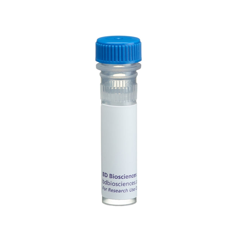
BD Pharmingen™ Purified Mouse anti-EGF Receptor (pY845)

BD Pharmingen™ Purified Mouse anti-EGF Receptor (pY845)

Regulatory Status Legend
Any use of products other than the permitted use without the express written authorization of Becton, Dickinson and Company is strictly prohibited.
Preparation And Storage
Product Notices
- Since applications vary, each investigator should titrate the reagent to obtain optimal results.
- Caution: Sodium azide yields highly toxic hydrazoic acid under acidic conditions. Dilute azide compounds in running water before discarding to avoid accumulation of potentially explosive deposits in plumbing.
- Sodium azide is a reversible inhibitor of oxidative metabolism; therefore, antibody preparations containing this preservative agent must not be used in cell cultures nor injected into animals. Sodium azide may be removed by washing stained cells or plate-bound antibody or dialyzing soluble antibody in sodium azide-free buffer. Since endotoxin may also affect the results of functional studies, we recommend the NA/LE (No Azide/Low Endotoxin) antibody format, if available, for in vitro and in vivo use.
- Please refer to www.bdbiosciences.com/us/s/resources for technical protocols.
Epidermal Growth Factor (EGF) elicits a variety of cellular responses that are initiated by EGF Receptor (EGFR) binding and activation of intrinsic tyrosine kinase activity. EGFR, also known as ErbB1 or HER1, is a member of the ErbB class of receptor protein tyrosine kinases. It has an extracellular ligand-binding domain, a single transmembrane region, and a cytoplasmic region containing a protein tyrosine kinase domain and a c-terminal regulatory domain with many phosphorylation sites. Following ligand binding, EGFR forms homodimers and heterodimers with ErbB2. Specific C-terminal tyrosine residues are then autophosphorylated and, in turn, bind to adaptor proteins, kinases, or protein tyrosine phosphatases. In addition, c-Src-dependent phosphorylations at other sites, such as tyrosine 845 (Y845) in the kinase domain, regulate the receptor's kinase activity. Inappropriate expression or mutations of EGFR and/or deregulation of its signaling pathways are associated with many types of cancer, making EGFR a promising target for cancer therapies.
The 12A3 monoclonal antibody recognizes the phosphorylated Y845 in the protein tyrosine kinase domain of human EGFR.
Development References (7)
-
Biscardi JS, Maa ME, Tice DA, Cox ME, Leu TH, Parsons SJ. c-Src-mediated phosphorylation of the epidermal growth factor receptor on Tyr845 and Tyr1101 is associated with modulation of receptor function. J Biol Chem. 1999; 274(12):8335-8343. (Biology).
-
Boerner JL, Demory ML, Silva C, Parsons SJ. Phosphorylation of Y845 on the epidermal growth factor receptor mediates binding to the mitochondrial protein cytochrome c oxidase subunit II. Mol Cell Biol. 2004; 24(16):7059-7071. (Biology).
-
Hristozova T, Konschak R, Budach V, Tinhofer I. A simple multicolor flow cytometry protocol for detection and molecular characterization of circulating tumor cells in epithelial cancers. Cytometry A. 2012; 81A(6):489-495. (Clone-specific: Flow cytometry). View Reference
-
Mendelsohn J, Baselga J. Status of epidermal growth factor receptor antagonists in the biology and treatment of cancer. J Clin Oncol. 2003; 21(14):2787-2799. (Biology).
-
Moro L, Dolce L, Cabodi S, et al. Integrin-induced epidermal growth factor (EGF) receptor activation requires c-Src and p13Cas and leads to phosphorylation of specific EGF receptor tyrosines. J Biol Chem. 2002; 277(11):9405-9414. (Biology).
-
Olayioye MA, Neve RM, Lane HA, Nynes NE. The ErbB signaling network: receptor heterodimerization in development and cancer. EMBO J. 2000; 19(13):3159-3167. (Biology).
-
Wu W, Graves LM, Gill GN, Parsons SJ, Samet JM. Src-dependent phosphorylation of the epidermal growth factor receptor on tyrosine 845 is required for zinc-induced Ras activation. J Biol Chem. 2002; 277(27):24252-24257. (Biology).
Please refer to Support Documents for Quality Certificates
Global - Refer to manufacturer's instructions for use and related User Manuals and Technical data sheets before using this products as described
Comparisons, where applicable, are made against older BD Technology, manual methods or are general performance claims. Comparisons are not made against non-BD technologies, unless otherwise noted.
For Research Use Only. Not for use in diagnostic or therapeutic procedures.
Report a Site Issue
This form is intended to help us improve our website experience. For other support, please visit our Contact Us page.
