
Preparation And Storage
Recommended Assay Procedures
BD® CompBeads can be used as surrogates to assess fluorescence spillover (compensation). When fluorochrome conjugated antibodies are bound to BD® CompBeads, they have spectral properties very similar to cells. However, for some fluorochromes there can be small differences in spectral emissions compared to cells, resulting in spillover values that differ when compared to biological controls. It is strongly recommended that when using a reagent for the first time, users compare the spillover on cells and BD® CompBeads to ensure that BD® CompBeads are appropriate for your specific cellular application.
Product Notices
- Since applications vary, each investigator should titrate the reagent to obtain optimal results.
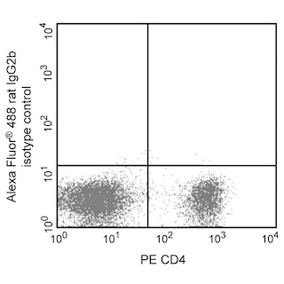
- An isotype control should be used at the same concentration as the antibody of interest.
- Caution: Sodium azide yields highly toxic hydrazoic acid under acidic conditions. Dilute azide compounds in running water before discarding to avoid accumulation of potentially explosive deposits in plumbing.
- Alexa Fluor® 488 fluorochrome emission is collected at the same instrument settings as for fluorescein isothiocyanate (FITC).
- For fluorochrome spectra and suitable instrument settings, please refer to our Multicolor Flow Cytometry web page at www.bdbiosciences.com/colors.
- Species cross-reactivity detected in product development may not have been confirmed on every format and/or application.
- Please refer to www.bdbiosciences.com/us/s/resources for technical protocols.
- This product is provided under an intellectual property license between Life Technologies Corporation and BD Businesses. The purchase of this product conveys to the buyer the non-transferable right to use the purchased amount of the product and components of the product in research conducted by the buyer (whether the buyer is an academic or for-profit entity). The buyer cannot sell or otherwise transfer (a) this product (b) its components or (c) materials made using this product or its components to a third party or otherwise use this product or its components or materials made using this product or its components for Commercial Purposes. Commercial Purposes means any activity by a party for consideration and may include, but is not limited to: (1) use of the product or its components in manufacturing; (2) use of the product or its components to provide a service, information, or data; (3) use of the product or its components for therapeutic, diagnostic or prophylactic purposes; or (4) resale of the product or its components, whether or not such product or its components are resold for use in research. For information on purchasing a license to this product for any other use, contact Life Technologies Corporation, Cell Analysis Business Unit Business Development, 29851 Willow Creek Road, Eugene, OR 97402, USA, Tel: (541) 465-8300. Fax: (541) 335-0504.
- Please refer to http://regdocs.bd.com to access safety data sheets (SDS).
- Alexa Fluor™ is a trademark of Life Technologies Corporation.
Companion Products


The M1/70 monoclonal antibody specifically binds to CD11b, also known as Integrin alpha M (Itgam or αM). CD11b is a 170-kDa type 1 transmembrane glycoprotein and belongs to the Integrin alpha chain family. CD11b serves as the alpha chain of the heterodimeric Mac-1 integrin (CD11b/CD18, αMβ2), also known as complement receptor 3 (CR3). Mac-1 mediates adhesion to ICAM-1 (CD54), ICAM-2 (CD102), fibrinogen and binding to C3bi. Mac-1 is expressed at varying levels on granulocytes, macrophages, myeloid-derived dendritic cells, natural killer cells, microglia, and B-1 B lymphocytes. Mac-1 expression is rapidly upregulated on neutrophils after activation, in the same time period that CD62L (L-selectin) is shed from the cell surface. The M1/70 antibody reportedly blocks cell adherence and C3bi binding but does not block cell-mediated lysis. Cross-reaction of the M1/70 antibody with CD11b expressed on human monocytes, polymorphonuclear leukocytes, and NK cells has been reported.
Development References (9)
-
Ault KA, Springer TA. Cross-reaction of a rat-anti-mouse phagocyte-specific monoclonal antibody (anti-Mac-1) with human monocytes and natural killer cells. J Immunol. 1981; 126(1):359-364. (Clone-specific). View Reference
-
Beller DI, Springer TA, Schreiber RD. Anti-Mac-1 selectively inhibits the mouse and human type three complement receptor. J Exp Med. 1982; 156(4):1000-1009. (Biology: Blocking). View Reference
-
Kishimoto TK, Jutila MA, Berg EL, Butcher EC. Neutrophil Mac-1 and MEL-14 adhesion proteins inversely regulated by chemotactic factors. Science. 1989; 245(4923):1238-1241. (Biology). View Reference
-
Lagasse E, Weissman IL. Flow cytometric identification of murine neutrophils and monocytes. J Immunol Methods. 1996; 197(1-2):139-150. (Methodology: Flow cytometry). View Reference
-
Lub M, van Kooyk Y, Figdor CG. Competition between lymphocyte function-associated antigen 1 (CD11a/CD18) and Mac-1 (CD11b/CD18) for binding to intercellular adhesion molecule-1 (CD54). J Leukoc Biol. 1996; 59(5):648-655. (Biology: Immunoprecipitation). View Reference
-
Sanchez-Madrid F, Simon P, Thompson S, Springer TA. Mapping of antigenic and functional epitopes on the alpha- and beta-subunits of two related mouse glycoproteins involved in cell interactions, LFA-1 and Mac-1. J Exp Med. 1983; 158(2):586-602. (Biology: Blocking, Immunoprecipitation, Western blot). View Reference
-
Springer T, Galfre G, Secher DS, Milstein C. Mac-1: a macrophage differentiation antigen identified by monoclonal antibody. Eur J Immunol. 1979; 9(4):301-306. (Clone-specific: Immunoprecipitation). View Reference
-
Springer T, Galfre G, Secher DS, Milstein C. Monoclonal xenogeneic antibodies to murine cell surface antigens: identification of novel leukocyte differentiation antigens. Eur J Immunol. 1978; 8(8):539-551. (Immunogen: Immunoprecipitation). View Reference
-
Springer TA, Davignon D, Ho MK, Kurzinger K, Martz E, Sanchez-Madrid F. LFA-1 and Lyt-2,3, molecules associated with T lymphocyte-mediated killing; and Mac-1, an LFA-1 homologue associated with complement receptor function. Immunol Rev. 1982; 68:171-195. (Biology: Blocking). View Reference
Please refer to Support Documents for Quality Certificates
Global - Refer to manufacturer's instructions for use and related User Manuals and Technical data sheets before using this products as described
Comparisons, where applicable, are made against older BD Technology, manual methods or are general performance claims. Comparisons are not made against non-BD technologies, unless otherwise noted.
For Research Use Only. Not for use in diagnostic or therapeutic procedures.