-
Reagents
- Flow Cytometry Reagents
-
Western Blotting and Molecular Reagents
- Immunoassay Reagents
-
Single-Cell Multiomics Reagents
- BD® AbSeq Assay
- BD Rhapsody™ Accessory Kits
- BD® Single-Cell Multiplexing Kit
- BD Rhapsody™ Targeted mRNA Kits
- BD Rhapsody™ Whole Transcriptome Analysis (WTA) Amplification Kit
- BD Rhapsody™ TCR/BCR Profiling Assays for Human and Mouse
- BD® OMICS-Guard Sample Preservation Buffer
- BD Rhapsody™ ATAC-Seq Assays
-
Functional Assays
-
Microscopy and Imaging Reagents
-
Cell Preparation and Separation Reagents
-
Training
- Flow Cytometry Basic Training
-
Product-Based Training
- BD FACSDiscover™ S8 Cell Sorter Product Training
- Accuri C6 Plus Product-Based Training
- FACSAria Product Based Training
- FACSCanto Product-Based Training
- FACSLyric Product-Based Training
- FACSMelody Product-Based Training
- FACSymphony Product-Based Training
- HTS Product-Based Training
- LSRFortessa Product-Based Training
- Advanced Training
-
- BD® AbSeq Assay
- BD Rhapsody™ Accessory Kits
- BD® Single-Cell Multiplexing Kit
- BD Rhapsody™ Targeted mRNA Kits
- BD Rhapsody™ Whole Transcriptome Analysis (WTA) Amplification Kit
- BD Rhapsody™ TCR/BCR Profiling Assays for Human and Mouse
- BD® OMICS-Guard Sample Preservation Buffer
- BD Rhapsody™ ATAC-Seq Assays
-
- BD FACSDiscover™ S8 Cell Sorter Product Training
- Accuri C6 Plus Product-Based Training
- FACSAria Product Based Training
- FACSCanto Product-Based Training
- FACSLyric Product-Based Training
- FACSMelody Product-Based Training
- FACSymphony Product-Based Training
- HTS Product-Based Training
- LSRFortessa Product-Based Training
- United States (English)
-
Change country/language
Old Browser
This page has been recently translated and is available in French now.
Looks like you're visiting us from {countryName}.
Would you like to stay on the current country site or be switched to your country?


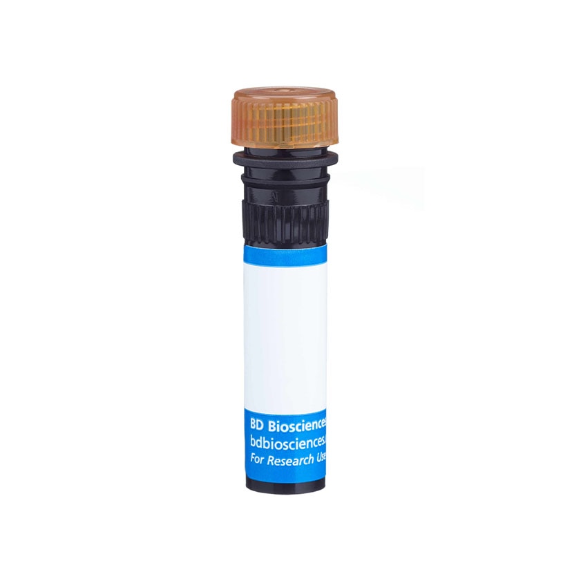

Flow cytometric analysis of IL-9 expressed in stimulated human CD4-positive T cells. Human peripheral blood mononuclear cells were stimulated in a tissue culture plate coated with NA/LE Mouse Anti-Human CD3 (Cat. No. 555329; 10 µg/ml, coated overnight at 4°C) and soluble NA/LE Mouse Anti-Human CD28 (Cat. No. 555725; 1 µg/ml) antibodies plus recombinant Human IL-2 (Cat. No. 554603; 10 ng/ml), IL-4 (Cat. No. 554605; 50 ng/ml), and TGF-β (Cat. No. 356039; 10 ng/ml) proteins and NA/LE Mouse Anti-Human IFN-γ (Cat. No. 554698; 10 µg/ml) antibody for 5 days. The cells were harvested and restimulated with PMA (Sigma P8139; 50 ng/ml) and ionomycin (Sigma I9657; 1 µg/ml) in the presence of BD GolgiStop™ Protein Transport Inhibitor (Cat. No. 554724) for 5 hours. The cells were then fixed and permeabilized using the BD Cytofix/Cytoperm™ Fixation/Permeablization Kit (Cat. No. 554714) followed by staining with PerCP-Cy™5.5 Mouse Anti-Human IL-9 (Cat. No. 561461) and Mouse Anti-Human CD4 (Cat. No. 555347) antibodies. Two-color flow cytometric dot plots showing IL-9 versus autofluorescence (PE channel) were derived from CD4 positive gated events with the forward and side light-scatter characteristics of viable lymphocytes. Flow cytometry was performed on a BD LSRII™ System.


BD Pharmingen™ PerCP-Cy™5.5 Mouse Anti-Human IL-9

Regulatory Status Legend
Any use of products other than the permitted use without the express written authorization of Becton, Dickinson and Company is strictly prohibited.
Preparation And Storage
Product Notices
- Source of all serum proteins is from USDA inspected abattoirs located in the United States.
- This reagent has been pre-diluted for use at the recommended Volume per Test. We typically use 1 × 10^6 cells in a 100-µl experimental sample (a test).
- An isotype control should be used at the same concentration as the antibody of interest.
- Please refer to www.bdbiosciences.com/us/s/resources for technical protocols.
- Cy is a trademark of Amersham Biosciences Limited. This conjugated product is sold under license to the following patents: US Patent Nos. 5,486,616; 5,569,587; 5,569,766; 5,627,027.
- Please observe the following precautions: Absorption of visible light can significantly alter the energy transfer occurring in any tandem fluorochrome conjugate; therefore, we recommend that special precautions be taken (such as wrapping vials, tubes, or racks in aluminum foil) to prevent exposure of conjugated reagents, including cells stained with those reagents, to room illumination.
- Caution: Sodium azide yields highly toxic hydrazoic acid under acidic conditions. Dilute azide compounds in running water before discarding to avoid accumulation of potentially explosive deposits in plumbing.
- PerCP-Cy5.5–labelled antibodies can be used with FITC- and R-PE–labelled reagents in single-laser flow cytometers with no significant spectral overlap of PerCP-Cy5.5, FITC, and R-PE fluorescence.
- PerCP-Cy5.5 is optimized for use with a single argon ion laser emitting 488-nm light. Because of the broad absorption spectrum of the tandem fluorochrome, extra care must be taken when using dual-laser cytometers, which may directly excite both PerCP and Cy5.5™. We recommend the use of cross-beam compensation during data acquisition or software compensation during data analysis.
- For fluorochrome spectra and suitable instrument settings, please refer to our Multicolor Flow Cytometry web page at www.bdbiosciences.com/colors.
- This product is subject to proprietary rights of Amersham Biosciences Corp. and Carnegie Mellon University and made and sold under license from Amersham Biosciences Corp. This product is licensed for sale only for research. It is not licensed for any other use. If you require a commercial license to use this product and do not have one return this material, unopened to BD Biosciences, 10975 Torreyana Rd, San Diego, CA 92121 and any money paid for the material will be refunded.
Companion Products

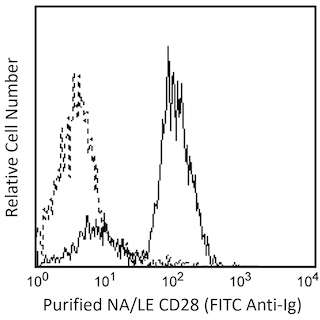
The MH9A3 monoclonal antibody specifically binds to human interleukin-9 (IL-9). Human IL-9 is a multifunctional cytokine and a member of the type I cytokine (hematopoietin) family that includes IL-2, IL-4, IL-7, IL-15 and IL-21. This cytokine is encoded by the IL9 gene that is resident on chromosome 5q31.1. IL-9 is expressed by activated CD4-positive T helper cells, by some transformed T cells and by eosinophils, mast cells and neutrophils. IL-9 induces the proliferation, differentiation, and effector function of various cell types including T lymphocytes, B lymphocytes, mast cells, eosinophils, neutrophils, hematopoietic cells and epithelial cells. It potentiates the interleukin-4-induced IgM, IgG and IgE responses by human B lymphocytes. IL-9 has been implicated in human allergic disorders such as asthma and malignancies such as Hodgkin's disease. IL-9 exerts its biological activities through binding to the surface IL-9 receptor (IL-9R) complex comprised of the IL-9R alpha subunit (IL-9Rα; CD129) and the common cytokine receptor gamma subunit (γc; CD132). IL-9 signaling through its receptor includes activation of the Janus kinases 1 and 3 ( JAK1 and JAK3) and activation of Signal transducer and activator of transcription 1, 3 and 5 factors (STAT1, STAT3 and STAT5).

Development References (9)
-
Demoulin JB, Van Roost E, Stevens M, Groner B, Renauld JC. Distinct roles for STAT1, STAT3, and STAT5 in differentiation gene induction and apoptosis inhibition by interleukin-9. J Biol Chem. 1999; 274:25855-25861. (Biology). View Reference
-
Dugas B, Renauld JC, Pène J, et al. Interleukin-9 potentiates the interleukin-4-induced immunoglobulin (IgG, IgM and IgE) production by normal human B lymphocytes. Eur J Immunol. 1993 July; 23(7):1687-1692. (Biology). View Reference
-
Houssiau FA, Schandene L, Stevens M. A cascade of cytokines is responsible for IL-9 expression in human T cells. Involvement of IL-2, IL-4, and IL-10. J Immunol. 1995; 154(6):2624-2630. (Biology). View Reference
-
Jenmalm MC, Van Snick J, Cormont F, Salman B. Allergen-induced Th1 and Th2 cytokine secretion in relation to specific allergen sensitization and atopic symptoms in children. Clin Exp Allergy. 2001 October; 31(10):1528-1535. (Clone-specific: ELISA). View Reference
-
Knoops L, Renauld JC. IL-9 and its receptor: from signal transduction to tumorigenesis. Growth Factors. 2004; 22:207-215. (Biology). View Reference
-
Merz H, Houssiau FA, Orscheschek K, et al. Interleukin-9 expression in human malignant lymphomas: unique association with Hodgkin's disease and large cell anaplastic lymphoma. Blood. 1991 September; 78(5):1311-1317. (Biology). View Reference
-
Renauld JC. New insights into the role of cytokines in asthma. J Clin Pathol. 2001 August; 54(8):577-589. (Biology). View Reference
-
Soler D, Chapman TR, Poisson LR. CCR8 expression identifies CD4 memory T cells enriched for FOXP3+ regulatory and Th2 effector lymphocytes. J Immunol. 2006; 177(10):6940-6951. (Biology). View Reference
-
Soroosh P, Doherty TA. Th9 and allergic disease. Immunology. 2009; 127(4):450-458. (Biology). View Reference
Please refer to Support Documents for Quality Certificates
Global - Refer to manufacturer's instructions for use and related User Manuals and Technical data sheets before using this products as described
Comparisons, where applicable, are made against older BD Technology, manual methods or are general performance claims. Comparisons are not made against non-BD technologies, unless otherwise noted.
For Research Use Only. Not for use in diagnostic or therapeutic procedures.
Report a Site Issue
This form is intended to help us improve our website experience. For other support, please visit our Contact Us page.