

Preparation And Storage
Product Notices
- Please refer to www.bdbiosciences.com/us/s/resources for technical protocols.
- Since applications vary, each investigator should titrate the reagent to obtain optimal results.
- Caution: Sodium azide yields highly toxic hydrazoic acid under acidic conditions. Dilute azide compounds in running water before discarding to avoid accumulation of potentially explosive deposits in plumbing.
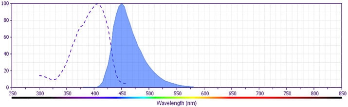
- BD Horizon V450 has a maximum absorption of 406 nm and maximum emission of 450 nm. Before staining with this reagent, please confirm that your flow cytometer is capable of exciting the fluorochrome and discriminating the resulting fluorescence.
- Pacific Blue™ is a trademark of Molecular Probes, Inc., Eugene, OR.
Companion Products

.png?imwidth=320)
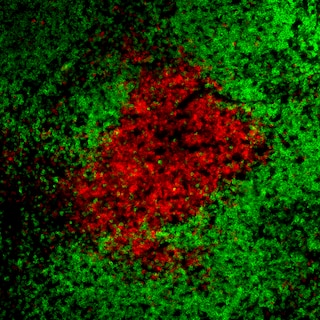
The 500A2 monoclonal antibody specifically binds to the 25-kDa ε chain of the T-cell receptor-associated CD3 complex expressed on mouse thymocytes, mature T lymphocytes, and NKT cells. Plate-bound and soluble forms of the 500A2 antibody can activate T cells in vitro. Activation of a mouse T-cell clone by the 500A2 antibody can be blocked by Fab fragments of the GK1.5 anti-CD4 antibody. This suggests that the 500A2 antibody may bind an epitope on CD3e close to a site at which CD4 associates with the T-cell receptor. The 500A2 antibody reportedly does not to crossreact with rat leukocytes.
The antibody is conjugated to BD Horizon™ V450, which has been developed for use in multicolor flow cytometry experiments and is available exclusively from BD Biosciences. It is excited by the Violet laser Ex max of 406 nm and has an Em Max at 450 nm. Conjugates with BD Horizon™ V450 can be used in place of Pacific Blue™ conjugates.
Development References (5)
-
Allison JP, Havran WL, Poenie M, et al. Expression and function of CD3 on murine thymocytes. In: Kappler J, Davis M, ed. The T-Cell Receptor, UCLA Symposia, 73rd Edition. Los Angeles: 1988:33-45.
-
Havran WL, Poenie M, Kimura J, Tsien R, Weiss A, Allison JP. Expression and function of the CD3-antigen receptor on murine CD4+8+ thymocytes. Nature. 1987; 330(6144):170-173. (Clone-specific). View Reference
-
Kubo RT, Born W, Kappler JW, Marrack P, Pigeon M. Characterization of a monoclonal antibody which detects all murine alpha beta T cell receptors. J Immunol. 1989; 142(8):2736-2742. (Methodology). View Reference
-
Ortaldo JR, Winkler-Pickett R, Mason AT, Mason LH. The Ly-49 family: regulation of cytotoxicity and cytokine production in murine CD3+ cells. J Immunol. 1998; 160(1):1158-1165. (Clone-specific). View Reference
-
Portoles P, Rojo J, Golby A, et al . Monoclonal antibodies to murine CD3 epsilon define distinct epitopes, one of which may interact with CD4 during T cell activation. J Immunol. 1989; 142(12):4169-4175. (Clone-specific). View Reference
Please refer to Support Documents for Quality Certificates
Global - Refer to manufacturer's instructions for use and related User Manuals and Technical data sheets before using this products as described
Comparisons, where applicable, are made against older BD Technology, manual methods or are general performance claims. Comparisons are not made against non-BD technologies, unless otherwise noted.
For Research Use Only. Not for use in diagnostic or therapeutic procedures.