
Preparation And Storage
Recommended Assay Procedures
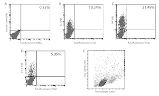
Immunofluorescent Staining and Flow Cytometry: The APC-conjugated MP6-XT22 antibody can be used for multicolor immunofluorescent staining and flow cytometric analyses to identify and enumerate TNF-producing cells within mixed cell populations (see figure). For optimal immunofluorescent staining with flow cytometric analysis, this anti-cytokine antibody should be titrated (≤ 0.5 µg mAb/million cells). For specific methodology, please visit our web site, www.bdbiosciences.com, and go to the protocols section or the chapter on intracellular staining in the Immune Function Handbook.
A useful control for demonstrating specificity of staining is either of the following: 1) pre-block the conjugated MP6-XT22 antibody with ligand (e.g., recombinant mouse TNF; Cat. No. 554589) prior to staining, or 2) pre-block the fixed/permeabilized cells with unlabeled MP6-XT22 antibody (Cat. No. 554416) prior to staining. The staining technique and use of blocking controls are described in detail by C. Prussin and D. Metcalfe. A suitable rat IgG1 isotype control for assessing the level of background staining on paraformaldehyde-fixed/saponin-permeabilized mouse cells is APC-R3-34 (Cat. No. 554686). Isotype controls should be used at a comparable concentration to the antibody of interest.
ELISA Capture: The purified MP6-XT22 antibody (Cat. No. 554416) is useful as a capture antibody for a sandwich ELISA for measuring mouse TNF protein levels.
WB: The MP6-XT22 antibody has been reported to be useful for Western blotting. Please note that this application is not routinely tested at BD Biosciences Pharmingen.
Product Notices
- Since applications vary, each investigator should titrate the reagent to obtain optimal results.
- Please refer to www.bdbiosciences.com/us/s/resources for technical protocols.
- For fluorochrome spectra and suitable instrument settings, please refer to our Multicolor Flow Cytometry web page at www.bdbiosciences.com/colors.
- Caution: Sodium azide yields highly toxic hydrazoic acid under acidic conditions. Dilute azide compounds in running water before discarding to avoid accumulation of potentially explosive deposits in plumbing.
Companion Products

.png?imwidth=320)
The MP6-XT22 antibody specifically binds to mouse Tumor Necrosis Factor (TNF, also known as TNF-α). TNF is produced by many activated cell types including monocytes, macrophages, astrocytes, granulocytes, mast cells, T and B lymphocytes, NK cells, keratinocytes, fibroblasts, adipocytes, and certain tumor cells. Activated cells express type II transmembrane TNF glycoproteins that associate as homotrimeric complexes. After enzymatic cleavage, the extracellular regions of membrane TNF are shed as soluble homotrimers. TNF is a potent multifunctional cytokine that can exert regulatory and cytotoxic effects on a wide range of normal lymphoid and non-lymphoid cells and tumor cells. Although TNF serves as a primary mediator in protective immune responses against microbial and viral pathogens, it can also drive systemic pathophysiologic responses including septic shock, cachexia and autoimmune diseases. Mouse TNF exerts its biological activities by binding and signaling through cell surface membrane Type I and Type II TNF Receptors (aka, TNFRI/CD120a and TNFRII/CD120b, respectively).

Development References (4)
-
Abrams J. Immunoenzymetric assay of mouse and human cytokines using NIP-labeled anti-cytokine antibodies. Curr Protoc Immunol. 2001; 1:6.20-6.21. (Clone-specific). View Reference
-
Abrams JS, Roncarolo MG, Yssel H, Andersson U, Gleich GJ, Silver JE. Strategies of anti-cytokine monoclonal antibody development: immunoassay of IL-10 and IL-5 in clinical samples. Immunol Rev. 1992; 127:5-24. (Clone-specific). View Reference
-
Litton MJ, Sander B, Murphy E, O'Garra A, Abrams JS. Early expression of cytokines in lymph nodes after treatment in vivo with Staphylococcus enterotoxin B. J Immunol Methods. 1994; 175(1):47-58. (Clone-specific). View Reference
-
Prussin C, Metcalfe DD. Detection of intracytoplasmic cytokine using flow cytometry and directly conjugated anti-cytokine antibodies. J Immunol Methods. 1995; 188(1):117-128. (Methodology). View Reference
Please refer to Support Documents for Quality Certificates
Global - Refer to manufacturer's instructions for use and related User Manuals and Technical data sheets before using this products as described
Comparisons, where applicable, are made against older BD Technology, manual methods or are general performance claims. Comparisons are not made against non-BD technologies, unless otherwise noted.
For Research Use Only. Not for use in diagnostic or therapeutic procedures.