Skip to main content Skip to navigation
BV711 Mouse Anti-Human IL-2
Two color flow cytometric analysis of IL-2 expression by activated human peripheral blood lymphocytes. Human peripheral blood mononuclear cells were stimulated for 5 h with Phorbol 12-Myristate 13-Acetate (PMA, Sigma P-8139; 50 ng/ml) and Calcium Ionophore A23187 (Sigma C-9275; 1 μ g/ml), in the presence of BD GolgiStop™ Protein Transport Inhibitor (containing Monensin) (Cat. No. 554724). The cells were fixed with BD Cytofix/Cytoperm™ Fixation and Permeabilization Solution (Cat. No. 554722), washed and permeabilized with BD Perm/Wash™ Buffer (Cat. No. 554723), and then stained with FITC Mouse Anti-Human CD3 antibody (Cat No. 555332/561806/561807) and either BD Horizon™ BV711 Mouse IgG1, κ Isotype Control (Cat No. 563044, Left Panel) or BD Horizon™ BV711 Mouse Anti-Human IL-2 antibody (Cat No. 563946, Right Panel) by using BD Biosciences Intracellular Cytokine Staining Protocol. The two-color flow cytometric dot plots show the correlated expression of IL-2 (or Ig Isotype control staining) versus CD3 and were derived from gated events with the forward and side light-scatter characteristics of intact lymphocytes. Flow cytometric analysis was performed using a BD™ LSR II Flow Cytometer System.
Product Details
Down Arrow Up Arrow


BD Horizon™
IL2; Interleukin-2; T-cell growth factor; TCGF
Human (QC Testing)
Mouse BALB/c IgG1, κ
Human IL-2 protein
Intracellular staining (flow cytometry) (Routinely Tested)
5 µl
3558
AB_2738501
Aqueous buffered solution containing BSA and ≤0.09% sodium azide.
RUO


Preparation And Storage

Store undiluted at 4°C and protected from prolonged exposure to light. Do not freeze. The monoclonal antibody was purified from tissue culture supernatant or ascites by affinity chromatography. The antibody was conjugated with BD Horizon™ BV711 under optimum conditions, and unconjugated antibody and free BD Horizon™ BV711 were removed.

Recommended Assay Procedures

For optimal and reproducible results, BD Horizon Brilliant Stain Buffer should be used anytime two or more BD Horizon Brilliant dyes are used in the same experiment.  Fluorescent dye interactions may cause staining artifacts which may affect data interpretation.  The BD Horizon Brilliant Stain Buffer was designed to minimize these interactions.  More information can be found in the Technical Data Sheet of the BD Horizon Brilliant Stain Buffer (Cat. No. 563794/566349) or the BD Horizon Brilliant Stain Buffer Plus (Cat. No. 566385).

Product Notices

  1. This reagent has been pre-diluted for use at the recommended Volume per Test. We typically use 1 × 10^6 cells in a 100-µl experimental sample (a test).
  2. An isotype control should be used at the same concentration as the antibody of interest.
  3. Source of all serum proteins is from USDA inspected abattoirs located in the United States.
  4. Caution: Sodium azide yields highly toxic hydrazoic acid under acidic conditions. Dilute azide compounds in running water before discarding to avoid accumulation of potentially explosive deposits in plumbing.
  5. Alexa Fluor® is a registered trademark of Molecular Probes, Inc., Eugene, OR.
  6. For fluorochrome spectra and suitable instrument settings, please refer to our Multicolor Flow Cytometry web page at www.bdbiosciences.com/colors.
  7. BD Horizon Brilliant Violet 711 is covered by one or more of the following US patents: 8,110,673; 8,158,444; 8,227,187; 8,455,613; 8,575,303; 8,354,239.
  8. BD Horizon Brilliant Stain Buffer is covered by one or more of the following US patents: 8,110,673; 8,158,444; 8,575,303; 8,354,239.
  9. Cy is a trademark of GE Healthcare.
  10. Please refer to www.bdbiosciences.com/us/s/resources for technical protocols.
563946 Rev. 2
Antibody Details
Down Arrow Up Arrow
5344.111

The 5344.111 antibody specifically binds to the multifunctional cytokine, human interleukin-2 (IL-2). IL-2 is produced by activated T cells and has multiple functions that can affect the growth, proliferation and differentiation of many different target cell types including T cells, B cells, NK cells, monocytes and macrophages. The immunogen used to generate the 5344.111 hybridoma was affinity-purified, recombinant human IL-2 protein.

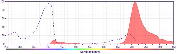
The antibody was conjugated to BD Horizon BV711 which is part of the BD Horizon Brilliant™ Violet family of dyes. This dye is a tandem fluorochrome of BD Horizon BV421 with an Ex Max of 405-nm and an acceptor dye with an Em Max at 711-nm.  BD Horizon BV711 can be excited by the violet laser and detected in a filter used to detect Cy™5.5 / Alexa Fluor® 700-like dyes (eg, 712/20-nm filter).  Due to the excitation and emission characteristics of the acceptor dye, there may be moderate spillover into the Alexa Fluor® 700 and PerCP-Cy5.5 detectors.  However, the spillover can be corrected through compensation as with any other dye combination.

563946 Rev. 2
Format Details
Down Arrow Up Arrow
BV711
The BD Horizon Brilliant Violet™ 711 (BV711) Dye is part of the BD Horizon Brilliant Violet™ family of dyes. This tandem fluorochrome is comprised of a BV421 donor with an excitation maximum (Ex Max) of 407-nm and an acceptor dye with an emission maximum (Em Max) at 713-nm. BV711, driven by BD innovation, is designed to be excited by the violet laser (405-nm) and detected using an optical filter centered near 710-nm (e.g., a 712/20-nm bandpass filter). The acceptor dye can be excited by the Red (628–640-nm) laser resulting in cross-laser excitation and fluorescence spillover. Please ensure that your instrument’s configurations (lasers and optical filters) are appropriate for this dye.
altImg
BV711
Violet 405 nm
407 nm
713 nm
563946 Rev.2
Citations & References
Down Arrow Up Arrow

Development References (4)

  1. Liao W, Lin JX, Leonard WJ. IL-2 family cytokines: new insights into the complex roles of IL-2 as a broad regulator of T helper cell differentiation. Curr Protoc Immunol. 2011; 23(5):598-604. (Biology). View Reference
  2. Malek TR. The biology of interleukin-2. Annu Rev Immunol. 2008; 26:453-479. (Biology). View Reference
  3. Prussin C, Metcalfe DD. Detection of intracytoplasmic cytokine using flow cytometry and directly conjugated anti-cytokine antibodies. J Immunol Methods. 1995; 188(1):117-128. (Methodology: Flow cytometry). View Reference
  4. Waldmann TA. The biology of interleukin-2 and interleukin-15: implications for cancer therapy and vaccine design. Nat Rev Immunol. 2006; 6(8):595-601. (Biology). View Reference
View All (4) View Less
563946 Rev. 2

Please refer to Support Documents for Quality Certificates


Global - Refer to manufacturer's instructions for use and related User Manuals and Technical data sheets before using this products as described


Comparisons, where applicable, are made against older BD Technology, manual methods or are general performance claims.  Comparisons are not made against non-BD technologies, unless otherwise noted.

For Research Use Only. Not for use in diagnostic or therapeutic procedures.