

Preparation And Storage
Product Notices
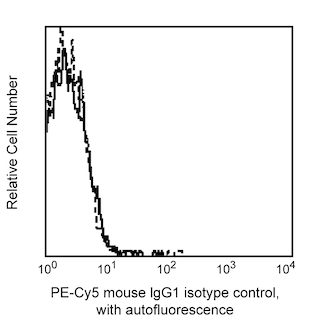
- This reagent has been pre-diluted for use at the recommended Volume per Test. We typically use 1 × 10^6 cells in a 100-µl experimental sample (a test).
- An isotype control should be used at the same concentration as the antibody of interest.
- Caution: Sodium azide yields highly toxic hydrazoic acid under acidic conditions. Dilute azide compounds in running water before discarding to avoid accumulation of potentially explosive deposits in plumbing.
- Source of all serum proteins is from USDA inspected abattoirs located in the United States.
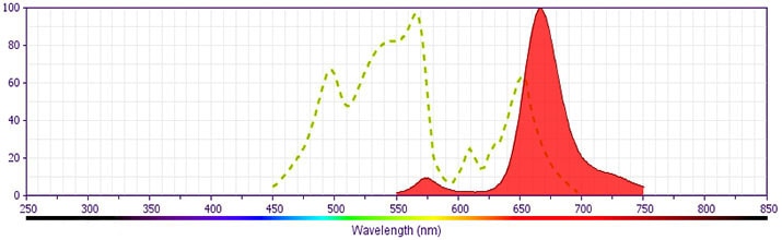
- PE-Cy5 is optimized for use with a single argon ion laser emitting 488-nm light. Because of the broad absorption spectrum of the PE-Cy5 tandem fluorochrome, extra care must be taken when using dual-laser cytometers which may directly excite both PE and Cy5™.
- PE-Cy5 is a tandem fluorochrome composed of R-phycoerythrin (PE), which is excited by the 488 nm light of an Argon ion laser and serves as an energy donor, coupled to the cyanine dye Cy5, which acts as an energy acceptor and fluoresces at 670 nm. BD Biosciences Pharmingen has maximized the fluorochrome energy transfer in PE-Cy5, thus maximizing its fluorescence emission intensity, minimizing residual emission from PE, and minimizing lot-to-lot variation.
- PE-Cy5 tandem fluorochromes have been reported to bind some classes of human macrophages and granulocytes via Fc receptors, and PE has been reported to bind to mouse B lymphocytes via Fc receptors. Preincubation of mouse leukocytes with Mouse BD Fc Block™ purified anti-mouse CD16/CD32 mAb 2.4G2 can reduce the non-specific binding of PE-Cy5-conjugated reagents to mouse B cells. However, PE-Cy5 conjugated reagents should not be used to stain splenocytes of SJL, NOD, and MRL mice as B lymphocytes and/or other leukocytes have been reported to non-specifically stain regardless of the use of Mouse BD Fc Block™ (the CD72c complex has been implicated for PE-Cy5 binding in these strains). Reagents conjugated to PE, PerCP, PerCP-Cy5.5, APC, and APC-Cy7 tandem fluorochrome can be used on leukocytes from these mouse strains.
- Cy is a trademark of GE Healthcare.
- Species cross-reactivity detected in product development may not have been confirmed on every format and/or application.
- Please observe the following precautions: Absorption of visible light can significantly alter the energy transfer occurring in any tandem fluorochrome conjugate; therefore, we recommend that special precautions be taken (such as wrapping vials, tubes, or racks in aluminum foil) to prevent exposure of conjugated reagents, including cells stained with those reagents, to room illumination.
- For fluorochrome spectra and suitable instrument settings, please refer to our Multicolor Flow Cytometry web page at www.bdbiosciences.com/colors.
- Please refer to www.bdbiosciences.com/us/s/resources for technical protocols.
The 2331 (FUN-1) monoclonal antibody specifically recognizes a 75 kDa transmembrane cell surface protein, CD86 (B70/B7-2), expressed primarily on monocytes, dendritic cells and activated B cells. Competitive binding assays demonstrate that, while both 2331 (FUN-1) and IT2.2 (Anti-CD86) antibodies specifically recognize the same molecule, they react with different epitopes. CD86 is a ligand for CD28 and CTLA-4 and plays an important role in costimulation of T cells in primary immune response. The 2331 (FUN-1) antibody blocks the costimulatory activity of CD86 when tested in functional studies.
Development References (5)
-
Azuma H, Uno Y, Shigekiyo T, Saito S. Congenital plasminogen deficiency caused by a Ser572 to Pro mutation. Blood. 1993; 82(2):475-480. (Biology). View Reference
-
Engel P, Gribben JG, Freeman GJ, et al. The B7-2 (B70) costimulatory molecule expressed by monocytes and activated B lymphocytes is the CD86 differentiation antigen. Blood. 1994; 84(5):1402-1407. (Biology). View Reference
-
Engel P, Wagner N, Tedder TF. CD86 Workshop Report. In: Schlossman SF. Stuart F. Schlossman .. et al., ed. Leucocyte typing V : white cell differentiation antigens : proceedings of the fifth international workshop and conference held in Boston, USA, 3-7 November, 1993. Oxford: Oxford University Press; 1995:703-705.
-
Nozawa Y, Wachi E, Tominaga K, Abe M, Wakasa H. A novel monoclonal antibody (FUN-1) identifies an activation antigen in cells of the B-cell lineage and Reed-Sternberg cells. J Pathol. 1993; 169(3):309-315. (Clone-specific). View Reference
-
Yang XF, Chen Z, Wormsley SB. Nashville: American Society of Hematology; 1994.
Please refer to Support Documents for Quality Certificates
Global - Refer to manufacturer's instructions for use and related User Manuals and Technical data sheets before using this products as described
Comparisons, where applicable, are made against older BD Technology, manual methods or are general performance claims. Comparisons are not made against non-BD technologies, unless otherwise noted.
For Research Use Only. Not for use in diagnostic or therapeutic procedures.