

Preparation And Storage
Recommended Assay Procedures
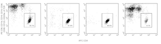
Peripheral Blood Mononuclear Cells (PBMC) are labeled with BD IMag™ Anti-Human CD25 Magnetic Particles - DM according to the Magnetic Labeling and Separation Protocol. This labeled cell suspension is then placed within the magnetic field of the BD IMag™ Cell Separation Magnet. Labeled cells migrate toward the magnet (positive fraction), leaving the unlabeled cells in suspension so they can be drawn off (negative fraction). The tube is then removed from the magnetic field for the resuspension of the positive fraction. The separation is repeated twice to increase the purity of the positive fraction. The magnetic separation steps are diagrammed in the Separation Flow Chart. After the positive fraction is washed, it can be further evaluated in downstream applications. The small size of the BD IMag™ particles allows the positive fraction to be further evaluated in downstream applications, such as by flow cytometry.
MAGNETIC LABELING AND SEPARATION PROTOCOL
1. Dilute BD IMag™ Buffer (10X) (Cat. No. 552362) 1:10 with sterile distilled water or prepare 1X BD IMag™ buffer by supplementing Phosphate Buffered Saline with 0.5% BSA, 2 mM EDTA, and 0.09% sodium azide. Store at 4°C.
2. Prepare PBMC from anti-coagulated human blood, preferably by density gradient centrifugation using Ficoll-Paque™.
Optional: If Treg cells are desired, enrich the CD4 T lymphocytes by using the BD IMag™ Human CD4 T Lymphocyte Enrichment Set - DM (Cat. No. 557939).
3. Count the cells, wash them with an excess volume of 1X BD IMag™ buffer, and carefully aspirate all the supernatant.
4. Vortex the BD IMag™ Anti-Human CD25 Magnetic Particles - DM thoroughly, and add 50 µl of particles for every 10^7 total cells.
5. MIX THOROUGHLY. Incubate at room temperature for 30 minutes.†
6. Bring the BD IMag™-particle labeling volume up to 1-8 x 10^7 cells/mL with 1X BD IMag™ buffer, and immediately place the tube on the Cell Separation Magnet. Incubate for 8 - 10 minutes.
7. With the tube on the Cell Separation Magnet, carefully aspirate off the supernatant. This supernatant contains the negative fraction.
8. Remove the tube from the Cell Separation Magnet, and add 1X BD IMag™ buffer to the same volume as in Step 6. Gently resuspend cells by pipetting up and down, and return the tube to the Cell Separation Magnet for another 2 - 4 minutes.
9. With the tube on the Cell Separation Magnet, carefully aspirate off the supernatant and discard.
10. Repeat Steps 8 and 9.
11. After the final wash step, resuspend the positive fraction in an appropriate buffer or medium, and proceed with desired downstream application(s).
NOTES:
* Hints for successful cell preparation:
- Draw the blood into a tube containing EDTA.
- Remove the platelet rich plasma by centrifuging once at 220-240 × g.
- Wash 2-3 times in PBS after the density gradient separation.
- Remove clumps of cells and/or debris by passing the suspension through a 70 µm nylon cell strainer.
† Avoid nonspecific labeling by working quickly and adhering to recommended incubation times.
Product Notices
- Source of all serum proteins is from USDA inspected abattoirs located in the United States.
- Caution: Sodium azide yields highly toxic hydrazoic acid under acidic conditions. Dilute azide compounds in running water before discarding to avoid accumulation of potentially explosive deposits in plumbing.
- BD IMag™ particles are prepared from carboxy-functionalized magnetic particles which are manufactured by Skold Technology and are licensed under US patent number 7,169,618.
- Use of these products to measure activation antigens expressed on mononuclear cell subsets for the purpose of monitoring immunoregulatory status can fall under one or more claims of the following patents: US Patent Nos. 5,445,939, 5,656,446, 5,843,689; European Patent No. 319,543; Canadian Patent No. 1,296,622; Australian Patent No. 615,880; and Japanese Patent No. 2,769,156.
- Ficoll-Paque is a trademark of Amersham Biosciences Limited.
- Please refer to www.bdbiosciences.com/us/s/resources for technical protocols.
Companion Products
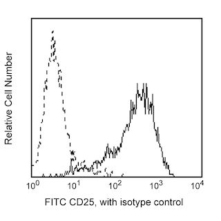
BD IMag™ Anti-Human CD25 Magnetic Particles - DM are magnetic nanoparticles that have monoclonal antibody conjugated to their surfaces. These particles are optimized for the positive selection or depletion of CD25-bearing leukocytes using the BD IMag™ Cell Separation Magnet. CD25 is the 55 kDa α chain of the IL-2 receptor that is expressed on activated B and T lymphocytes, which may also include regulatory T-cell (Treg cells). For enrichment of Treg cells, depletion of erythrocytes, platelets, monocytes, granulocytes and non-CD4 T lymphocytes is first recommended using the BD IMag™ Human CD4 T Lymphocyte Enrichment Set - DM, followed by the positive selection of the CD25+ population.
Development References (2)
-
Curotto de Lafaille MA, Lafaille JJ. CD4(+) regulatory T cells in autoimmunity and allergy. Curr Opin Immunol. 2002; 14(6):771-778. (Biology). View Reference
-
Knapp W. W. Knapp .. et al., ed. Leucocyte typing IV : white cell differentiation antigens. Oxford New York: Oxford University Press; 1989:1-1182.
Please refer to Support Documents for Quality Certificates
Global - Refer to manufacturer's instructions for use and related User Manuals and Technical data sheets before using this products as described
Comparisons, where applicable, are made against older BD Technology, manual methods or are general performance claims. Comparisons are not made against non-BD technologies, unless otherwise noted.
For Research Use Only. Not for use in diagnostic or therapeutic procedures.