
Preparation And Storage
Recommended Assay Procedures
Note: For best staining results, cell samples should be stained and maintained at 4-8°C before flow cytometric analysis.
BD® CompBeads can be used as surrogates to assess fluorescence spillover (Compensation). When fluorochrome conjugated antibodies are bound to BD® CompBeads, they have spectral properties very similar to cells. However, for some fluorochromes there can be small differences in spectral emissions compared to cells, resulting in spillover values that differ when compared to biological controls. It is strongly recommended that when using a reagent for the first time, users compare the spillover on cells and BD® CompBeads to ensure that BD® CompBeads are appropriate for your specific cellular application.
Product Notices
- This reagent has been pre-diluted for use at the recommended Volume per Test. We typically use 1 × 10^6 cells in a 100-µl experimental sample (a test).
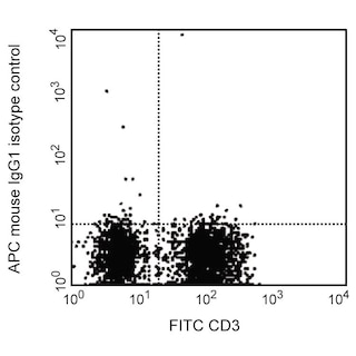
- An isotype control should be used at the same concentration as the antibody of interest.
- Source of all serum proteins is from USDA inspected abattoirs located in the United States.
- Caution: Sodium azide yields highly toxic hydrazoic acid under acidic conditions. Dilute azide compounds in running water before discarding to avoid accumulation of potentially explosive deposits in plumbing.
- This APC-conjugated reagent can be used in any flow cytometer equipped with a dye, HeNe, or red diode laser.
- For fluorochrome spectra and suitable instrument settings, please refer to our Multicolor Flow Cytometry web page at www.bdbiosciences.com/colors.
- Please refer to www.bdbiosciences.com/us/s/resources for technical protocols.
- Please refer to http://regdocs.bd.com to access safety data sheets (SDS).
Companion Products


The BerH8 monoclonal antibody specifically binds to CD30, a 120 kDa type I transmembrane glycoprotein expressed on stimulated T and B cells. CD30 is an activation marker initially identified to be expressed on Reed-Sternberg cells. It is also expressed on a few extrafollicular T and B cells located at the rim of germinal centers. CD30 serves as a cytokine receptor. It belongs to the nerve growth factor receptor/tumor necrosis factor receptor (NGFR/TNFR) superfamily and is also known as TNFRSF8. CD30 interaction with CD30 ligand (CD30L/CD153/TNFSF8) can mediate signals for proliferation, apoptosis and cytotoxicity of lymphoid cells.

Development References (5)
-
Beverly PCL. Activation antigens: new and previously defined clusters. In: McMichael AJ. A.J. McMichael .. et al., ed. Leucocyte typing III : white cell differentiation antigens. Oxford New York: Oxford University Press; 1987:516-524.
-
Bowen MA, Olsen KJ, Cheng L, Avila D, Podack ER. Functional effects of CD30 on a large granular lymphoma cell line, YT. Inhibition of cytotoxicity, regulation of CD28 and IL-2R, and induction of homotypic aggregation. J Immunol. 1993; 151(11):5896-5906. (Clone-specific: Immunohistochemistry). View Reference
-
Franke AC, Jung D, Ellis TM. Characterization of the CD30L binding domain on the human CD30 molecule using anti-CD30 antibodies. Hybridoma. 2000; 19(1):43-48. (Clone-specific: Blocking, Flow cytometry). View Reference
-
Schlossman SF. Stuart F. Schlossman .. et al., ed. Leucocyte typing V : white cell differentiation antigens : proceedings of the fifth international workshop and conference held in Boston, USA, 3-7 November, 1993. Oxford: Oxford University Press; 1995.
-
Schwarting R, Stein H. Cluster report CD30. In: Knapp W. W. Knapp .. et al., ed. Leucocyte typing IV : white cell differentiation antigens. Oxford New York: Oxford University Press; 1989:419-422.
Please refer to Support Documents for Quality Certificates
Global - Refer to manufacturer's instructions for use and related User Manuals and Technical data sheets before using this products as described
Comparisons, where applicable, are made against older BD Technology, manual methods or are general performance claims. Comparisons are not made against non-BD technologies, unless otherwise noted.
For Research Use Only. Not for use in diagnostic or therapeutic procedures.