
Description
Rat Monocyte Chemoattractant Protein (MCP-1) is a member of the C-C chemokine superfamily and a homolog of human MCP-1. MCP-1 is expressed by a variety of cell types including monocyte/macrophages, endothelial cells and mesangial cells.3 MCP-1 has chemoattractant activity for monocytes, lymphocytes and basophils but is not active for neutrophils. Recombinant rat MCP-1 is a highly glycosylated protein ranging in size from 27 - 30 kD as measured by SDS-PAGE analysis. Furthermore, there is evidence from experimental animal models that MCP-1 can suppress tumor formation by attracting monocytes to the tumor site. Recombinant rat MCP-1 (MN 555110) is supplied as a frozen liquid comprised of 0.22 μm sterile-filtered aqueous buffered solution containing glycerol and 1.0 mg/ml bovine serum albumin, with no preservatives. Recombinant rat MCP-1 is ≥ 95% pure as determined by SDS-PAGE, and an absorbance assay based on the Beers-Lambert law. The endotoxin level is ≤ 0.1 ng per µg of rat MCP-1, as measured in a chromogenic LAL assay.
Preparation And Storage
Recommended Assay Procedures
Upon initial thawing, recombinant rat MCP-1 (MN 555110) should be aliquoted into polypropylene microtubes and frozen at -80°C for future use. Alternatively, the product can be diluted in sterile neutral buffer containing not less than 0.5 - 10 mg/mL carrier protein, such as human or bovine albumin, aliquoted and stored at -80°C. For in vitro biological assay use, carrier-protein concentrations of 0.5 - 1 mg/mL are recommended. For use as an ELISA standard, carrier-protein concentrations of 5 - 10 mg/mL are recommended. Failure to add carrier protein or store at indicated temperatures may result in a loss of activity. Carrier proteins should be pre-screened for possible affects in each investigator's experimental system. Carrier proteins may have an undesired influence on experimental results due to toxicity, high endotoxin levels or possible blocking activity.
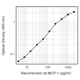
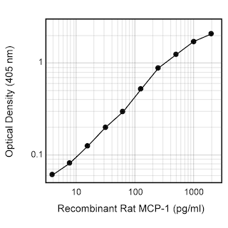
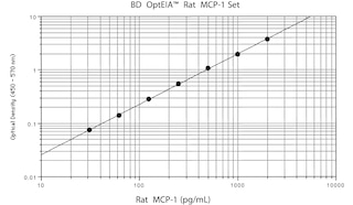
ELISA Standard: Recombinant rat MCP-1 (MN 555110) can be useful as a quantitative standard for measuring rat MCP-1 protein levels using sandwich ELISA with the purified C4 antibody (Cat. No. 555072) as a capture antibody and the biotinylated B4 antibody (Cat. No. 555074) as the detection antibody. To obtain linear standard curves, investigators may want to consider using doubling dilutions of recombinant rat MCP-1 from 2000-5 pg/mL to be included for each ELISA plate. For measuring rat MCP-1 in serum or plasma, investigators are highly encouraged to use the BD OptEIA™ Rat MCP-1 Set (Cat. No. 555130).
Bioassay: Investigators are advised that the Bioassay application is not routinely tested for this material and are highly encouraged to both titrate this material and include appropriate controls in relevant experiments. An activity range encompassing an ED50 = 60 - 600 ng/mL has previously been reported using THP-1 as indicator cells utilizing a calcium flux assay.
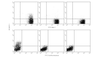
Ligand Blocking Control for Immunofluorescent Staining of Cytokines: Recombinant rat MCP-1 can be used as a blocking control to demonstrate the specificity of MCP-1 staining by the PE-conjugated format (Cat. No.554443) of the 2H5 Anti-Rat MCP-1 antibody. Investigators are advised that this blocking application is not routinely tested for this material. The use of staining controls for the immunofluorescent staining and flow cytometric analysis of cytokine producing cells has been previously described (Prussin et al.).
Product Notices
- Since applications vary, each investigator should titrate the reagent to obtain optimal results.
- Source of all serum proteins is from USDA inspected abattoirs located in the United States.
- Please refer to www.bdbiosciences.com/us/s/resources for technical protocols.
Companion Products
Development References (5)
-
Capsoni F, Minonzio F, Ongari AM, Zanussi C. A new simplified single-filter assay for 'in vitro' evaluation of chemotaxis of 51Cr-labeled polymorphonuclear leukocytes. J Immunol Methods. 1989; 120(1):125-131. (Methodology). View Reference
-
Haelens A, Wuyts A, Proost P, Struyf S, Opdenakker G, van Damme J.. Leukocyte migration and activation by murine chemokines. Immunobiology. 1996; 195(4-5):499-521. (Biology). View Reference
-
Prussin C, Metcalfe DD. Detection of intracytoplasmic cytokine using flow cytometry and directly conjugated anti-cytokine antibodies. J Immunol Methods. 1995; 188(1):117-128. (Methodology). View Reference
-
Rollins BJ, Sunday ME. Suppression of tumor formation in vivo by expression of the JE gene in malignant cells.. Mol Cell Biol. 1991; 11(6):3125-3131. (Biology). View Reference
-
Yoshimura T, Takeya M, Takahashi K. Molecular cloning of rat monocyte chemoattractant protein-1 (MCP-1) and its expression in rat spleen cells and tumor cell lines. Biochem Biophys Res Commun. 1991; 174(2):504-509. (Biology). View Reference
Please refer to Support Documents for Quality Certificates
Global - Refer to manufacturer's instructions for use and related User Manuals and Technical data sheets before using this products as described
Comparisons, where applicable, are made against older BD Technology, manual methods or are general performance claims. Comparisons are not made against non-BD technologies, unless otherwise noted.
For Research Use Only. Not for use in diagnostic or therapeutic procedures.