Skip to main content Skip to navigation
BV510 Mouse anti-BrdU
Two-color flow cytometric analysis of DNA synthesis by TK-1 cells. BrdU labeling and staining of cells was performed using the procedure and reagents, including BrdU, fixation and permeabilization buffers and 7-AAD Staining Solution from the BD Pharmingen™ FITC BrdU Flow Kit (Cat. No. 559619). Cells from the TK-1 cell line were either labeled with 50 μM BrdU for 1 hour (Left Panel) or were not labeled (Right Panel). The permeabilized cells were stained with BD Horizon™ BV510 Mouse Anti-BrdU antibody (Cat. No. 563445) followed by staining with the 7-AAD solution. Two-color flow cytometric dot plots showing the correlated expression patterns of 7-AAD (total cellular DNA) versus incorporated BrdU (representing newly-synthesized DNA) were derived from gated events with the forward and side light-scatter characteristics of intact cells. Flow cytometric analysis was performed using a BD™ LSR II System.
Product Details
Down Arrow Up Arrow


BD Horizon™
5-bromo-2'-deoxyuridine, 5-Bromouracil deoxyriboside, BUdR
Mouse (QC Testing), Human, Rat (Tested in Development)
Mouse IgG1, κ
Intracellular staining (flow cytometry) (Routinely Tested)
5 µl
AB_2738210
Aqueous buffered solution containing BSA and ≤0.09% sodium azide.
RUO


Preparation And Storage

Store undiluted at 4°C and protected from prolonged exposure to light. Do not freeze. The monoclonal antibody was purified from tissue culture supernatant or ascites by affinity chromatography. The antibody was conjugated with BD Horizon™ BV510 under optimum conditions, and unconjugated antibody and free BD Horizon™ BV510 were removed.

Recommended Assay Procedures

For detailed protocol information please visit http://www.bdbiosciences.com/resources/protocols/brdu_detection.jsp

Product Notices

  1. Source of all serum proteins is from USDA inspected abattoirs located in the United States.
  2. An isotype control should be used at the same concentration as the antibody of interest.
  3. This reagent has been pre-diluted for use at the recommended Volume per Test. We typically use 1 × 10^6 cells in a 100-µl experimental sample (a test).
  4. Please refer to www.bdbiosciences.com/us/s/resources for technical protocols.
  5. Caution: Sodium azide yields highly toxic hydrazoic acid under acidic conditions. Dilute azide compounds in running water before discarding to avoid accumulation of potentially explosive deposits in plumbing.
  6. For fluorochrome spectra and suitable instrument settings, please refer to our Multicolor Flow Cytometry web page at www.bdbiosciences.com/colors.
  7. Brilliant Violet™ 510 is a trademark of Sirigen.
  8. Species cross-reactivity detected in product development may not have been confirmed on every format and/or application.
563445 Rev. 1
Antibody Details
Down Arrow Up Arrow
3D4

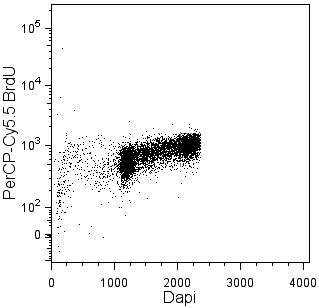
Bromodeoxyuridine (BrdU) is an analog of thymidine that can be incorporated into newly synthesized DNA by cells entering and progressing through the DNA synthesis (S) phase of the cell cycle.  The amount of incorporated BrdU depends on the amount of time that the cells are exposed to BrdU (pulse time), the rate of cell division, and whether the cells are in early, mid, or late S phase. Investigators can identify cycling cells in an asynchronous cell population and determine cell cycle kinetics by detecting incorporated BrdU.

The 3D4 monoclonal antibody reacts with BrdU, but not other nucleotides, in single-stranded DNA. Random cleavage (nicking) of cellular DNA with DNase I permits the binding of the antibody to incorporated BrdU.

The antibody was conjugated to BD Horizon™ BV510 which is part of the BD Horizon™ Brilliant Violet™ family of dyes. With an Ex Max of 405-nm and Em Max at 510-nm, BD Horizon™ BV510 can be excited by the violet laser and detected in the BD Horizon™ V500 (525/50-nm) filter set. BD Horizon™ BV510 conjugates are useful for the detection of dim markers off the violet laser.

563445 Rev. 1
Format Details
Down Arrow Up Arrow
BV510
The BD Horizon Brilliant Violet™ 510 (BV510) Dye is part of the BD Horizon Brilliant Violet™ family of dyes. This polymer-technology based dye with an excitation maximum (Ex Max) at 327-nm / 405-nm and an emission maximum (Em Max) at 512-nm. BV510, driven by BD innovation, is designed to be excited by the violet laser (405-nm) and detected using an optical filter centered near 510-nm (e.g., a 525/50 bandpass filter). The dye can be excited by the UV (355-nm) laser resulting in cross-laser excitation and spillover. Please ensure that your instrument’s configurations (lasers and optical filters) are appropriate for this dye.
altImg
BV510
Violet 405 nm
327 nm, 405 nm
512 nm
563445 Rev.1
Citations & References
Down Arrow Up Arrow

Development References (3)

  1. Dolbeare F, Gratzner H, Pallavicini MG, Gray JW. Flow cytometric measurement of total DNA content and incorporated bromodeoxyuridine. Proc Natl Acad Sci U S A. 1983; 80(18):5573-5577. (Methodology: Immunofluorescence). View Reference
  2. Keren DF, Hanson CA, Hurtubise PE. David F. Keren, Curtis A. Hanson, Paul E. Hurtubise., ed. Flow cytometry and clinical diagnosis. Chicago: ASCP Press; 1994:1-676.
  3. Miltenburger HG, Sachse G, Schliermann M. S-phase cell detection with a monoclonal antibody. Dev Biol Stand. 1987; 66:91-99. (Clone-specific: Immunofluorescence).
563445 Rev. 1

Please refer to Support Documents for Quality Certificates


Global - Refer to manufacturer's instructions for use and related User Manuals and Technical data sheets before using this products as described


Comparisons, where applicable, are made against older BD Technology, manual methods or are general performance claims.  Comparisons are not made against non-BD technologies, unless otherwise noted.

For Research Use Only. Not for use in diagnostic or therapeutic procedures.