-
Training
- Flow Cytometry Basic Training
-
Product-Based Training
- BD FACSDiscover™ S8 Cell Sorter Product Training
- Accuri C6 Plus Product-Based Training
- FACSAria Product Based Training
- FACSCanto Product-Based Training
- FACSLyric Product-Based Training
- FACSMelody Product-Based Training
- FACSymphony Product-Based Training
- HTS Product-Based Training
- LSRFortessa Product-Based Training
- Advanced Training
-
- BD FACSDiscover™ S8 Cell Sorter Product Training
- Accuri C6 Plus Product-Based Training
- FACSAria Product Based Training
- FACSCanto Product-Based Training
- FACSLyric Product-Based Training
- FACSMelody Product-Based Training
- FACSymphony Product-Based Training
- HTS Product-Based Training
- LSRFortessa Product-Based Training
- United States (English)
-
Change country/language
Old Browser
This page has been recently translated and is available in French now.
Looks like you're visiting us from {countryName}.
Would you like to stay on the current country site or be switched to your country?


Analysis of Lck in human peripheral blood lymphocytes. Human whole blood was lysed and fixed with 1X BD Phosflow™ Lyse/Fix Buffer (Cat. No. 558049) for 10-15 minutes at 37ºC, then permeabilized (BD Phosflow™ Perm Buffer II, Cat. No. 558052) on ice for 30 minutes, and then stained with either Alexa Fluor® 647 Mouse anti-Lck (Cat. No. 558505, solid-line histogram) or Alexa Fluor® 647 Mouse IgG1 κ Isotype control (Cat. No. 557783, dashed-line histogram). Flow cytometry was performed on a BD FACSCalibur™ flow cytometry system. The figure displays lymphocytes that were selected by their scatter profile.


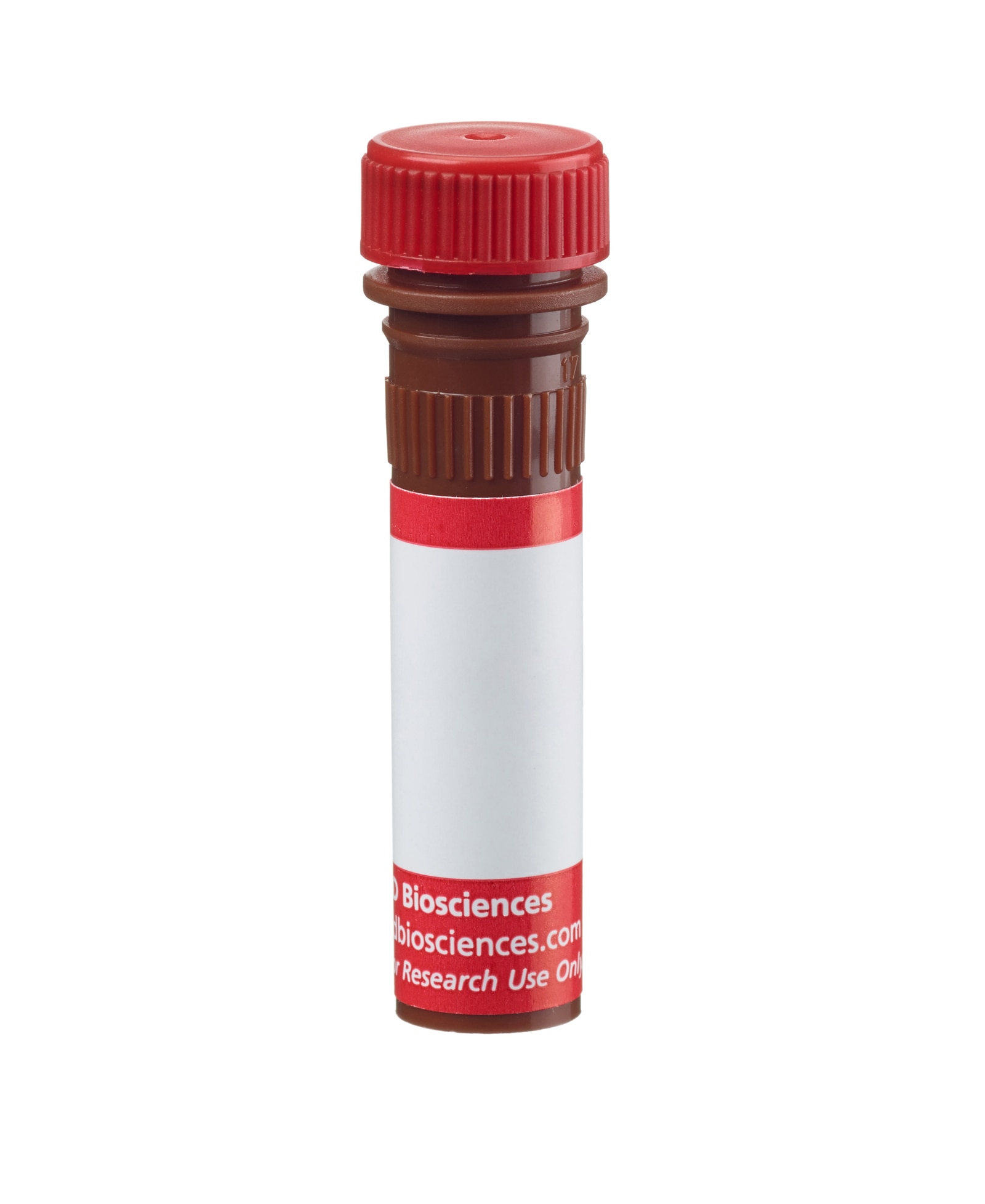
BD™ Phosflow Alexa Fluor® 647 Mouse anti-Lck

Regulatory Status Legend
Any use of products other than the permitted use without the express written authorization of Becton, Dickinson and Company is strictly prohibited.
Preparation And Storage
Product Notices
- This reagent has been pre-diluted for use at the recommended Volume per Test. We typically use 1 × 10^6 cells in a 100-µl experimental sample (a test).
- An isotype control should be used at the same concentration as the antibody of interest.
- Source of all serum proteins is from USDA inspected abattoirs located in the United States.
- Caution: Sodium azide yields highly toxic hydrazoic acid under acidic conditions. Dilute azide compounds in running water before discarding to avoid accumulation of potentially explosive deposits in plumbing.
- Alexa Fluor® 647 fluorochrome emission is collected at the same instrument settings as for allophycocyanin (APC).
- For fluorochrome spectra and suitable instrument settings, please refer to our Multicolor Flow Cytometry web page at www.bdbiosciences.com/colors.
- Alexa Fluor® is a registered trademark of Molecular Probes, Inc., Eugene, OR.
- The Alexa Fluor®, Pacific Blue™, and Cascade Blue® dye antibody conjugates in this product are sold under license from Molecular Probes, Inc. for research use only, excluding use in combination with microarrays, or as analyte specific reagents. The Alexa Fluor® dyes (except for Alexa Fluor® 430), Pacific Blue™ dye, and Cascade Blue® dye are covered by pending and issued patents.
- This product is provided under an intellectual property license between Life Technologies Corporation and BD Businesses. The purchase of this product conveys to the buyer the non-transferable right to use the purchased amount of the product and components of the product in research conducted by the buyer (whether the buyer is an academic or for-profit entity). The buyer cannot sell or otherwise transfer (a) this product (b) its components or (c) materials made using this product or its components to a third party or otherwise use this product or its components or materials made using this product or its components for Commercial Purposes. Commercial Purposes means any activity by a party for consideration and may include, but is not limited to: (1) use of the product or its components in manufacturing; (2) use of the product or its components to provide a service, information, or data; (3) use of the product or its components for therapeutic, diagnostic or prophylactic purposes; or (4) resale of the product or its components, whether or not such product or its components are resold for use in research. For information on purchasing a license to this product for any other use, contact Life Technologies Corporation, Cell Analysis Business Unit Business Development, 29851 Willow Creek Road, Eugene, OR 97402, USA, Tel: (541) 465-8300. Fax: (541) 335-0504.
- Species cross-reactivity detected in product development may not have been confirmed on every format and/or application.
- Please refer to http://regdocs.bd.com to access safety data sheets (SDS).
- Please refer to www.bdbiosciences.com/us/s/resources for technical protocols.
Companion Products


Lck is a member of the Src family of cytoplasmic protein-tyrosine kinases (PTKs) that is normally expressed exclusively in lymphoid cells, primarily T lymphocytes and NK cells. A low level of expression has been detected in B lymphocytes, but its function in B cells is unknown. Its expression in other leukocytes is not well defined. Members of the Src family have several common features: 1) unique N-terminal domains, 2) attachment to cellular membranes through a myristylated N-terminus, and 3) homologous SH2, SH3, and catalytic domains. The unique N-terminal domain of Lck interacts with the cytoplasmic tails of the CD4 and CD8 cell-surface glycoproteins of T lymphocytes, which recognize antigen presenting cells via their surface MHC class II and class I molecules, respectively. The catalytic activity of Lck is regulated by both kinases and phosphatases that control the phosphorylation states of two tyrosine residues that have opposing effects. Repression of Lck's catalytic activity occurs via phosphorylation at tyrosine 505 (Y505), located near the carboxy terminus. Phosphorylation of this tyrosine site is mediated by the Csk family of PTKs, and its dephosphorylation is mediated by the protein tyrosine phosphatase, CD45. When Lck is phosphorylated at this site, it assumes a folded tertiary structure which is enzymatically inactive. When CD45 dephosphorylates it at Y505, Lck is able to autophosphorylate its Y394, which leads to conformational changes in the catalytic domain that induce kinase activity. However, it has been observed that the inhibitory effect of the phosphorylated Y505 can be overcome by direct engagement of Lck's SH3 domain and that both Y394 and Y505 are phosphorylated together in cells activated by hydrogen peroxide. Activated Lck phosphorylates the ITAMs (Immunoreceptor-based Tyrosine Activation Motifs) of the T cell receptor (TCR) and thus is critical for activation and development of T lymphocytes. The interactions of Lck, Csk, CD45, CD4 or CD8, and TCR are only a small part of a complex immunoregulatory cascade that involves additional substrates for Csk and CD45, other enzymes, adhesion molecules, adaptor proteins, and specialized membrane microdomains.
The MOL 171 monoclonal antibody recognizes the 56- and 60-kDa forms of human Lck protein, regardless of phosphorylation status. It cross reacts with mouse Lck.
Development References (7)
-
Hardwick JS, Sefton BM. The activated form of the Lck tyrosine protein kinase in cells exposed to hydrogen peroxide is phosphorylated at both Tyr-394 and Tyr-505. J Biol Chem. 1997; 272:25429-25432. (Biology).
-
Holdorf AD, Lee K-H, Burack WR, Allen PM, Shaw AS. Regulation of Lck activity by CD4 and CD28 in the immunological synapse. Nat Immunol. 2002; 3(3):259-264. (Biology).
-
Johnson KG, Bromley SK, Dustin ML, Thomas ML. A supramolecular basis for CD45 tyrosine phosphatase regulation in sustained T cell activation. Proc Natl Acad Sci U S A. 2000; 97:10138-10143. (Biology).
-
Lee-Fruman KK, Collins TL, Burakoff SJ. Role of the Lck Src homology 2 and 3 domains in protein tyrosine phosphorylation. J Biol Chem. 1996; 271:25003-25010. (Biology).
-
Moroi Y, Koga Y, Nakamura K, Ohtsu M, Kimura G, Nomoto K. Accumulation of p60 lck in HTLV-I-transformed T cell lines detected by an anti-Lck monoclonal antibody, MOL 171. Jpn J Cancer Res. 1991; 82:909-915. (Immunogen).
-
Nakashima I, Pu M-Y, Hamaguchi M, et al. Pathway of signal delivery to murine thymocytes triggered by co-crosslinking CD3 and Thy-1 for cellular DNA fragmentation and growth inhibition. J Immunol. 1993; 151(7):3511-3520. (Clone-specific).
-
Veillette A, Latour S, Davidson D. Negative regulation of immunoreceptor signaling. Annu Rev Immunol. 2002; 20:669-707. (Biology).
Please refer to Support Documents for Quality Certificates
Global - Refer to manufacturer's instructions for use and related User Manuals and Technical data sheets before using this products as described
Comparisons, where applicable, are made against older BD Technology, manual methods or are general performance claims. Comparisons are not made against non-BD technologies, unless otherwise noted.
For Research Use Only. Not for use in diagnostic or therapeutic procedures.
Report a Site Issue
This form is intended to help us improve our website experience. For other support, please visit our Contact Us page.