

Preparation And Storage
Product Notices
- This reagent has been pre-diluted for use at the recommended Volume per Test. We typically use 1 × 10^6 cells in a 100-µl experimental sample (a test).
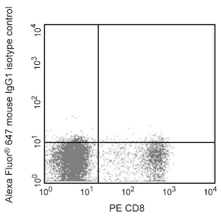
- An isotype control should be used at the same concentration as the antibody of interest.
- Source of all serum proteins is from USDA inspected abattoirs located in the United States.
- Caution: Sodium azide yields highly toxic hydrazoic acid under acidic conditions. Dilute azide compounds in running water before discarding to avoid accumulation of potentially explosive deposits in plumbing.
- The Alexa Fluor®, Pacific Blue™, and Cascade Blue® dye antibody conjugates in this product are sold under license from Molecular Probes, Inc. for research use only, excluding use in combination with microarrays, or as analyte specific reagents. The Alexa Fluor® dyes (except for Alexa Fluor® 430), Pacific Blue™ dye, and Cascade Blue® dye are covered by pending and issued patents.
- Alexa Fluor® is a registered trademark of Molecular Probes, Inc., Eugene, OR.
- Alexa Fluor® 647 fluorochrome emission is collected at the same instrument settings as for allophycocyanin (APC).
- For fluorochrome spectra and suitable instrument settings, please refer to our Multicolor Flow Cytometry web page at www.bdbiosciences.com/colors.
- Species cross-reactivity detected in product development may not have been confirmed on every format and/or application.
- Please refer to www.bdbiosciences.com/us/s/resources for technical protocols.
Companion Products


The 5D3-F7 monoclonal antibody specifically binds to human monocyte chemoattractant protein-1 (MCP-1), also known as CCL2 (C-C motif chemokine 2), Monocyte chemotactic and activating factor (MCAF), and Small-inducible cytokine A2 (SCYA2). MCP-1 is a 10-14 kDa glycoprotein member of the beta or CC family of chemokines. It expressed by monocytes, fibroblasts, endothelial cells and other cell types in response to IL-1, IL-6, TNF, and a variety of other stimuli. MCP-1 binds to and exerts its biological activity through G-protein coupled chemokine receptors including CCR2/CD192 and CCR4/CD194. It serves as a chemoattractant and activating factor for monocytes and other cell types including T cells, NK cells, and basophils.
MCP-1 is a member of the CC chemokine family and it is produced by monocytes, T lymphocytes, fibroblasts, endothelial cells, smooth muscle cells, keratinocytes and some tumors. Its production can be induced by LPS or IFN-γ. Clone 5D3-F7 also cross reacts with an intracellular component of LPS-stimulated (24 hours) peripheral blood monocytes of rhesus and cynomolgus macaque monkeys. The staining pattern observed on non-human primate monocytes is not as strong as that seen on normal human peripheral blood monocytes.
Development References (5)
-
Peri G, Milanese C, Matteucci C, et al. A new monoclonal antibody (5D3-F7) which recognizes human monocyte-chemotactic protein-1 but not related chemokines. Development of a sandwich ELISA and in situ detection of producing cells. J Immunol Methods. 1994; 174(1-2):249-257. (Immunogen: ELISA, Functional assay, Immunohistochemistry, Immunoprecipitation, Inhibition, Western blot). View Reference
-
Prussin C, Metcalfe DD. Detection of intracytoplasmic cytokine using flow cytometry and directly conjugated anti-cytokine antibodies. J Immunol Methods. 1995; 188(1):117-128. (Methodology). View Reference
-
Rollins BJ, Stier P, Ernst T, Wong GG. The human homolog of the JE gene encodes a monocyte secretory protein. Mol Cell Biol. 1989; 9(11):4687-4695. (Biology). View Reference
-
Vaddi K, Keller M, Newton RC. The chemokine factsbook. San Diego: Academic Press; 1997:205 p.
-
Yoshimura T, Yuhki N, Moore SK, Appella E, Lerman MI, Leonard EJ. Human monocyte chemoattractant protein-1 (MCP-1). Full-length cDNA cloning, expression in mitogen-stimulated blood mononuclear leukocytes, and sequence similarity to mouse competence gene JE. FEBS Lett. 1989; 244(2):487-493. (Biology). View Reference
Please refer to Support Documents for Quality Certificates
Global - Refer to manufacturer's instructions for use and related User Manuals and Technical data sheets before using this products as described
Comparisons, where applicable, are made against older BD Technology, manual methods or are general performance claims. Comparisons are not made against non-BD technologies, unless otherwise noted.
For Research Use Only. Not for use in diagnostic or therapeutic procedures.