
Preparation And Storage
Product Notices
- Since applications vary, each investigator should titrate the reagent to obtain optimal results.
- Please refer to www.bdbiosciences.com/us/s/resources for technical protocols.
- Caution: Sodium azide yields highly toxic hydrazoic acid under acidic conditions. Dilute azide compounds in running water before discarding to avoid accumulation of potentially explosive deposits in plumbing.
- Source of all serum proteins is from USDA inspected abattoirs located in the United States.
- For fluorochrome spectra and suitable instrument settings, please refer to our Multicolor Flow Cytometry web page at www.bdbiosciences.com/colors.
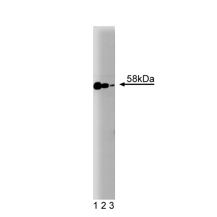
The transforming growth factor β (TGFβ)/activin/BMP family of growth factors plays a diverse and important role in growth, development, and differentiation. These growth factors act through their binding to heteromeric plasma membrane receptor protein kinases which, upon ligand binding, become activated and trigger an intracellular signaling cascade. Specifically, receptor activation induces the translocation of a set of conserved proteins named Smads (Sma- and Mad-related proteins) to the nucleus, resulting in gene activation. Smad2 is a ubiquitously expressed protein of 58 kDa that is phosphorylated and translocated to the nucleus in response to TGFβ, but not BMP. The overall response to TGFβ is growth inhibition. The Smad2 gene is located in chromosome 18q21.1 which is often absent in several human cancers. Furthermore, some missense mutations on the Smad2 gene were identified in colorectal carcinomas, suggesting Smad2 may function as a tumor suppressor in normal cells.
Investigators should note that potential crossreactivity to Smad3 is predicted based on sequence homology of the immunogen, Mouse Smad2 aa. 142-263. In addition, reactivity to mouse Smad2, using siRNA knockdown, has recently been described (Dzwonek et al.). Reactivity to canine Smad3 has also been reported using nuclear extracts (Lehman et al.).
Development References (7)
-
Dzwonek J, Preobrazhenska O, Cazzola S, et al. Smad3 is a key nonredundant mediator of transforming growth factor beta signaling in Nme mouse mammary epithelial cells. Mol Cancer Res. 2009; 7(8):1342-1353. (Clone-specific: Immunoprecipitation, Western blot). View Reference
-
Eppert K, Scherer SW, Ozcelik H, et al. MADR2 maps to 18q21 and encodes a TGFbeta-regulated MAD-related protein that is functionally mutated in colorectal carcinoma. Cell. 1996; 86(4):543-552. (Biology). View Reference
-
Hayes S, Chawla A, Corvera S. TGF beta receptor internalization into EEA1-enriched early endosomes: role in signaling to Smad2. J Cell Biol. 2002; 158(7):1239-1249. (Clone-specific: Immunofluorescence, Western blot). View Reference
-
Hocevar BA, Smine A, Xu XX, Howe PH. The adaptor molecule Disabled-2 links the transforming growth factor beta receptors to the Smad pathway. EMBO J. 2001; 20(11):2789-2801. (Clone-specific: Immunoprecipitation, Western blot). View Reference
-
Lechleider RJ, de Caestecker MP, Dehejia A, Polymeropoulos MH, Roberts AB. Serine phosphorylation, chromosomal localization, and transforming growth factor-beta signal transduction by human bsp-1. J Biol Chem. 1996; 271(30):17617-17620. (Biology). View Reference
-
Lehmann K, Janda E, Pierreux CE, et al. Raf induces TGFbeta production while blocking its apoptotic but not invasive responses: a mechanism leading to increased malignancy in epithelial cells. Genes Dev. 2000; 14(20):2610-2622. (Clone-specific: Gel shift, Western blot). View Reference
-
Luo Q, Nieves E, Kzhyshkowska J, Angeletti RH. Endogenous transforming growth factor-beta receptor-mediated Smad signaling complexes analyzed by mass spectrometry. Mol Cell Proteomics. 2006; 5(7):1245-1260. (Clone-specific: Immunoprecipitation, Western blot). View Reference
Please refer to Support Documents for Quality Certificates
Global - Refer to manufacturer's instructions for use and related User Manuals and Technical data sheets before using this products as described
Comparisons, where applicable, are made against older BD Technology, manual methods or are general performance claims. Comparisons are not made against non-BD technologies, unless otherwise noted.
For Research Use Only. Not for use in diagnostic or therapeutic procedures.