Preparation And Storage
Store vials at 2°C to 8°C. Conjugated forms should not be frozen. Protect from exposure to light. Each reagent is stable until the expiration date shown on the bottle label when stored as directed.
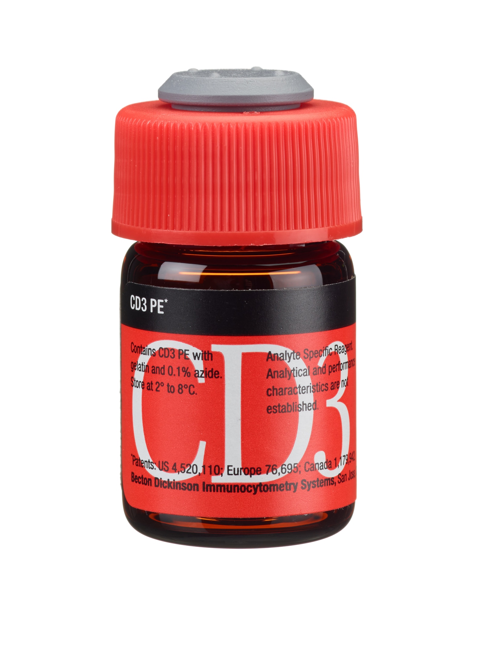
The CD3 antibody, clone SK7, is derived from the hybridization of NS-1 mouse myeloma cells with spleen cells isolated from BALB/c mice immunized with human thymocytes. The CD3 antibody reacts with the epsilon chain of the CD3 antigen/TCR complex. This complex is composed of at least six proteins that range in molecular weight from 20 to 30 kilodaltons (kDa). The antigen recognized by CD3 antibodies is noncovalently associated with either α/β or γ/δ TCR (70 to 90 kDa).

Development References (13)
-
Brenner M, Groh V, Porcelli S, et al. Knapp W, Dörken B, Gilks W, et al, ed. Leucocyte Typing IV: White Cell Differentiation Antigens. New York: Oxford University Press; 1989:1049-1053.
-
Centers for Disease Control. Update: universal precautions for prevention of transmission of human immunodeficiency virus, hepatitis B virus, and other bloodborne pathogens in healthcare settings. MMWR. 1988; 37:377-388. (Biology).
-
Clevers H, Alarcón B, Wileman T, Terhorst C. The T cell receptor/CD3 complex: a dynamic protein ensemble. Annual Rev Immunol. 1988; 6:629. (Biology).
-
Clinical and Laboratory Standards Institute. 2005. (Biology).
-
Garson JA, Beverley PCL, Coakham HB, Harper EJ. Monoclonal antibodies against human T lymphocytes label Purkinje neurones of many species. Nature. 1982; 298:375-377. (Biology).
-
Haynes BF. Summary of T-cell studies performed during the Second International Workshop and Conference on Human Leukocyte Differentiation Antigens. In: Reinherz EL. Ellis L. Reinherz .. et al., ed. Leukocyte typing II. New York: Springer-Verlag; 1986:3-30.
-
Kan EAR, Wang CY, Wang LC, Evans RL. Noncovalently bonded subunits of 22 and 28 kd are rapidly internalized by T cells reacted with Anti–Leu-4 antibody. J Immunol. 1983; 131:536-539. (Biology).
-
Kaneoka H, Perez-Rojas G, Sasasuki T, Benike CJ, Engleman EG. Human T lymphocyte proliferation induced by a pan-T monoclonal antibody (anti-Leu 4): heterogeneity of response is a function of monocytes. J Immunol. 1983; 131(1):158-164. (Biology). View Reference
-
Knowles RW. Immunochemical analysis of the T-cell–specific antigens. In: Reinherz EL. Ellis L. Reinherz .. et al., ed. Leukocyte typing II. New York: Springer-Verlag; 1986:259-288.
-
Lanier LL, Allison JP, Phillips JH. Correlation of cell surface antigen expression on human thymocytes by multi-color flow cytometric analysis: implications for differentiation. J Immunol. 1986; 137(8):2501-2507. (Biology). View Reference
-
Ledbetter JA, Evans RL, Lipinski M, Cunningham-Rundles C, Good RA, Herzenberg LA. Evolutionary conservation of surface molecules that distinguish T lymphocyte helper/inducer and cytotoxic/suppressor subpopulations in mouse and man. J Exp Med. 1981; 153(2):310-323. (Biology). View Reference
-
Reichert T, DeBruyere M, Deneys V, et al. Lymphocyte subset reference ranges in adult Caucasians. Clin Immunol Immunopathol. 1991; 60(2):190-208. (Biology). View Reference
-
van Dongen JJM, Krissansen GW, Wolvers-Tettero ILM, et al. Cytoplasmic expression of the CD3 antigen as a diagnostic marker for immature T-cell malignancies. Blood. 1988; 71:603-612. (Biology).
Please refer to Support Documents for Quality Certificates
Global - Refer to manufacturer's instructions for use and related User Manuals and Technical data sheets before using this products as described
Comparisons, where applicable, are made against older BD Technology, manual methods or are general performance claims. Comparisons are not made against non-BD technologies, unless otherwise noted.
Analyte Specific Reagent. Analytical and performance characteristics are not established.