
Preparation and Storage
推奨アッセイ手順
BD™ CompBeads can be used as surrogates to assess fluorescence spillover (Compensation). When fluorochrome conjugated antibodies are bound to BD CompBeads, they have spectral properties very similar to cells. However, for some fluorochromes there can be small differences in spectral emissions compared to cells, resulting in spillover values that differ when compared to biological controls. It is strongly recommended that when using a reagent for the first time, users compare the spillover on cells and BD CompBead to ensure that BD CompBeads are appropriate for your specific cellular application.
Product Notices
- Since applications vary, each investigator should titrate the reagent to obtain optimal results.
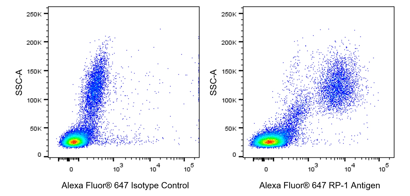
- An isotype control should be used at the same concentration as the antibody of interest.
- Caution: Sodium azide yields highly toxic hydrazoic acid under acidic conditions. Dilute azide compounds in running water before discarding to avoid accumulation of potentially explosive deposits in plumbing.
- The Alexa Fluor®, Pacific Blue™, and Cascade Blue® dye antibody conjugates in this product are sold under license from Molecular Probes, Inc. for research use only, excluding use in combination with microarrays, or as analyte specific reagents. The Alexa Fluor® dyes (except for Alexa Fluor® 430), Pacific Blue™ dye, and Cascade Blue® dye are covered by pending and issued patents.
- Alexa Fluor® is a registered trademark of Molecular Probes, Inc., Eugene, OR.
- Alexa Fluor® 647 fluorochrome emission is collected at the same instrument settings as for allophycocyanin (APC).
- For fluorochrome spectra and suitable instrument settings, please refer to our Multicolor Flow Cytometry web page at www.bdbiosciences.com/colors.
- This product is provided under an intellectual property license between Life Technologies Corporation and BD Businesses. The purchase of this product conveys to the buyer the non-transferable right to use the purchased amount of the product and components of the product in research conducted by the buyer (whether the buyer is an academic or for-profit entity). The buyer cannot sell or otherwise transfer (a) this product (b) its components or (c) materials made using this product or its components to a third party or otherwise use this product or its components or materials made using this product or its components for Commercial Purposes. Commercial Purposes means any activity by a party for consideration and may include, but is not limited to: (1) use of the product or its components in manufacturing; (2) use of the product or its components to provide a service, information, or data; (3) use of the product or its components for therapeutic, diagnostic or prophylactic purposes; or (4) resale of the product or its components, whether or not such product or its components are resold for use in research. For information on purchasing a license to this product for any other use, contact Life Technologies Corporation, Cell Analysis Business Unit Business Development, 29851 Willow Creek Road, Eugene, OR 97402, USA, Tel: (541) 465-8300. Fax: (541) 335-0504.
- Please refer to http://regdocs.bd.com to access safety data sheets (SDS).
- Please refer to www.bdbiosciences.com/us/s/resources for technical protocols.
関連製品


The RP-1 monoclonal antibody specifically recognizes the RP-1 Antigen. This cell surface marker is expressed on rat peritoneal and peripheral blood neutrophils. Amongst bone marrow cells, the RP-1 Antigen is expressed on band form and mature neutrophils but is not expressed on promyelocytes, myelocytes, and metamyelocytes. The RP-1 antibody does not bind to either rat monocytes, macrophages, eosinophils or to peritoneal neutrophils from mice, rabbits, guinea pigs, or to human peripheral blood neutrophils. Expression of the RP-1 Antigen on rat peritoneal neutrophils is enhanced by cellular stimulation with Phorbol 12-Myristate 13-Acetate (PMA) or Concanavalin A (ConA). Immunoprecipitation and SDS-PAGE analysis of non-treated and PMA-activated rat neutrophil membranes with the RP-1 antibody revealed two main bands of approximately 85 kDa. The RP-1 antibody is also known as the Mouse Anti-Rat Granulocytes antibody.
Development References (3)
-
Francis WR, Ireland RE, Spear AM, et al. Flow Cytometric Analysis of Hematopoietic Populations in Rat Bone Marrow. Impact of Trauma and Hemorrhagic Shock.. Cytometry A. 2019; 95(11):1167-1177. (Clone-specific: Flow cytometry, Fluorescence activated cell sorting). View Reference
-
Gotoh S, Itoh M, Fujii Y, Arai S, Sendo F. Enhancement of the expression of a rat neutrophil-specific cell surface antigen by activation with phorbol myristate acetate and concanavalin A. J Immunol. 1986; 137(2):643-650. (Immunogen: Flow cytometry, Immunoprecipitation, Radioimmunoassay). View Reference
-
Skrajnar S, Anzur Lasnik M, Bedina Zavec A. A flow cytometric method for determination of the blood neutrophil fraction in rats.. J Am Assoc Lab Anim Sci. 2009; 48(2):152-6. (Clone-specific: Flow cytometry). View Reference
Please refer to Support Documents for Quality Certificates
Global - Refer to manufacturer's instructions for use and related User Manuals and Technical data sheets before using this products as described
Comparisons, where applicable, are made against older BD Technology, manual methods or are general performance claims. Comparisons are not made against non-BD technologies, unless otherwise noted.
For Research Use Only. Not for use in diagnostic or therapeutic procedures.