
Preparation and Storage
推奨アッセイ手順
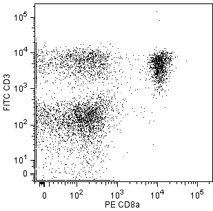
For flow cytometry of cell suspensions from peripheral lymphoid tissues, it is recommended that multicolor staining be performed to distinguish T lymphocytes from non-T-cells.
Product Notices
- Since applications vary, each investigator should titrate the reagent to obtain optimal results.
- An isotype control should be used at the same concentration as the antibody of interest.
- Caution: Sodium azide yields highly toxic hydrazoic acid under acidic conditions. Dilute azide compounds in running water before discarding to avoid accumulation of potentially explosive deposits in plumbing.
- For fluorochrome spectra and suitable instrument settings, please refer to our Multicolor Flow Cytometry web page at www.bdbiosciences.com/colors.
- Please refer to www.bdbiosciences.com/us/s/resources for technical protocols.
関連製品


.png?imwidth=320)
.png?imwidth=320)
The F23.1 antibody specifically reacts with the Vβ 8.1, Vβ 8.2, and Vβ 8.3 T-cell receptors (TCR) of mice having the b haplotype (e.g., A, AKR, BALB/c, CBA/Ca, CBA/J, C3H/He, C57BL, C58, DBA/1, DBA/2) of the Tcrb gene complex. The Tcrb-V8 subfamily gene loci are deleted in mice having the a (e.g., C57BR, C57L, SJL, SWR) or c (e.g., RIII) haplotype. Vβ 8.1 TCR-bearing T lymphocytes are clonally eliminated in mice expressing superantigen coded by Mtv-7 (Mls-1a, Mlsa) provirus (e.g., AKR, CBA/J, C58, DBA/2), and activation or elimination of Vβ 8.1 TCR-expressing T cells by this determinant is partially dependent upon presentation by I-E. Mtv-43 and/or exogenous MMTV-SW superantigens also cause incomplete elimination of Vβ 8.1 TCR-bearing T cells. In addition to expression on conventional T lymphocytes, Vβ 8.2 is the predominant β chain of the TCR on NK-T cells. Staphylococcal enterotoxin B, in association with antigen-presenting cells expressing I-A and/or I-E, stimulates lymphocytes bearing Vβ 8 TCR and selectively eliminates those T cells in vivo. Soluble and plate-bound F23.1 antibody activates Vβ 8 TCR-bearing T cells, soluble antibody blocks cytolysis mediated by Vβ 8 TCR-bearing cytotoxic T lymphocytes, and in vivo treatment of neonatal mice can arrest intrathymic maturation of Vβ 8 TCR-bearing T cells.

Development References (15)
-
Behlke MA, Chou HS, Huppi K, Loh DY. Murine T-cell receptor mutants with deletions of beta-chain variable region genes. Proc Natl Acad Sci U S A. 1986; 83(3):767-771. (Biology). View Reference
-
Behlke MA, Henkel TJ, Anderson SJ, et al. Expression of a murine polyclonal T cell receptor marker correlates with the use of specific members of the V beta 8 gene segment subfamily. J Exp Med. 1987; 165(1):257-262. (Clone-specific). View Reference
-
Bendelac A. Mouse NK1+ T cells. Curr Opin Immunol. 1995; 7(3):367-374. (Biology). View Reference
-
Haqqi TM, Banerjee S, Anderson GD, David CS. RIII S/J (H-2r). An inbred mouse strain with a massive deletion of T cell receptor V beta genes. J Exp Med. 1989; 169(6):1903-1909. (Biology). View Reference
-
Hodes RJ, Abe R. Mouse endogenous superantigens: Ms and Mls-like determinants encoded by mouse retroviruses.. Curr Protoc Immunol. 2001; Appendix 1:Appendix 1F. (Biology). View Reference
-
Hugo P, Kappler JW, Godfrey DI, Marrack PC. Thymic epithelial cell lines that mediate positive selection can also induce thymocyte clonal deletion. J Immunol. 1994; 52(3):1022-1031. (Biology). View Reference
-
Kappler JW, Staerz U, White J, Marrack PC. Self-tolerance eliminates T cells specific for Mls-modified products of the major histocompatibility complex. Nature. 1988; 332(6159):35-40. (Biology). View Reference
-
Kyewski BA, Schirrmacher V, Allison JP. Antibodies against the T cell receptor/CD3 complex interfere with distinct intra-thymic cell-cell interactions in vivo: correlation with arrest of T cell differentiation. Eur J Immunol. 1989; 19(5):857-863. (Clone-specific). View Reference
-
MacDonald HR, Baschieri S, Lees RK. Clonal expansion precedes anergy and death of V beta 8+ peripheral T cells responding to staphylococcal enterotoxin B in vivo. Eur J Immunol. 1991; 21(8):1963-1966. (Biology). View Reference
-
Mogil RJ, Radvanyi L, Gonzalez-Quintial R, et al. Fas (CD95) participates in peripheral T cell deletion and associated apoptosis in vivo. Int Immunol. 1995; 7(9):1451-1458. (Biology). View Reference
-
Renno T, Hahne M, Tschopp J, MacDonald HR. Peripheral T cells undergoing superantigen-induced apoptosis in vivo express B220 and upregulate Fas and Fas ligand. J Exp Med. 1996; 183(2):431-437. (Biology). View Reference
-
Staerz UD, Rammensee HG, Benedetto JD, Bevan MJ. Characterization of a murine monoclonal antibody specific for an allotypic determinant on T cell antigen receptor. J Immunol. 1985; 134(6):3994-4000. (Immunogen). View Reference
-
White J, Herman A, Pullen AM, Kubo R, Kappler JW, Marrack P. The V beta-specific superantigen staphylococcal enterotoxin B: stimulation of mature T cells and clonal deletion in neonatal mice. Cell. 1989; 56(1):27-35. (Biology). View Reference
-
Wolff CH, Hong SC, von Grafenstein H, Janeway CA Jr. TCR-CD4 and TCR-TCR interactions as distinctive mechanisms for the induction of increased intracellular calcium in T-cell signalling. J Immunol. 1993; 151(3):1337-1345. (Clone-specific). View Reference
-
Yagi J, Nakata M, Uchiyama T, et al. Superantigen-like properties of an antibody bispecific for MHC class II molecules and the V beta domain of the T cell antigen receptor. J Immunol. 1994; 152(8):3833-3841. (Clone-specific). View Reference
Please refer to Support Documents for Quality Certificates
Global - Refer to manufacturer's instructions for use and related User Manuals and Technical data sheets before using this products as described
Comparisons, where applicable, are made against older BD Technology, manual methods or are general performance claims. Comparisons are not made against non-BD technologies, unless otherwise noted.
For Research Use Only. Not for use in diagnostic or therapeutic procedures.