

Preparation and Storage
Product Notices
- This reagent has been pre-diluted for use at the recommended Volume per Test. We typically use 1 × 10^6 cells in a 100-µl experimental sample (a test).
- An isotype control should be used at the same concentration as the antibody of interest.
- Source of all serum proteins is from USDA inspected abattoirs located in the United States.
- Caution: Sodium azide yields highly toxic hydrazoic acid under acidic conditions. Dilute azide compounds in running water before discarding to avoid accumulation of potentially explosive deposits in plumbing.
- The Alexa Fluor®, Pacific Blue™, and Cascade Blue® dye antibody conjugates in this product are sold under license from Molecular Probes, Inc. for research use only, excluding use in combination with microarrays, or as analyte specific reagents. The Alexa Fluor® dyes (except for Alexa Fluor® 430), Pacific Blue™ dye, and Cascade Blue® dye are covered by pending and issued patents.
- Alexa Fluor® is a registered trademark of Molecular Probes, Inc., Eugene, OR.
- Alexa Fluor® 647 fluorochrome emission is collected at the same instrument settings as for allophycocyanin (APC).
- For fluorochrome spectra and suitable instrument settings, please refer to our Multicolor Flow Cytometry web page at www.bdbiosciences.com/colors.
- Please refer to http://regdocs.bd.com to access safety data sheets (SDS).
- Please refer to www.bdbiosciences.com/us/s/resources for technical protocols.
関連製品


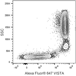
The MIH65.rMAb is a recombinant monoclonal antibody derived from MIH65 hybridoma cells. MIH65.rMAb specifically binds to human V-domain Ig suppressor of T cell activation (VISTA) like the conventional MIH65 antibody and performs like the MIH65 antibody when used to stain cells and analyze them by flow cytometry. VISTA is also known as B7-H5 (B7H5), PD-1H, DD1alpha (DD1α), chromosome 10 open reading frame 54 (C10orf54), Platelet Receptor Gi24 (GI24), or Stress induced secreted protein 1 (SISP1). VISTA is a 55-65 kDa single-pass, type I transmembrane glycoprotein that is encoded by VSIR (V-set immunoregulatory receptor) which belongs to the V-set domain containing family within the Ig superfamily. VISTA is primarily expressed on precursors and mature cells of the hematopoietic lineage including, monocytes, macrophages, neutrophils, dendritic cells (DC), T cells, regulatory T cells (Treg), and natural killer (NK) cells but not B cells. VISTA functions as an inhibitory regulator that can suppress T cell proliferation and cytokine production.
Development References (2)
-
Bharaj P, Chahar HS, Alozie OK, et al. Characterization of programmed death-1 homologue-1 (PD-1H) expression and function in normal and HIV infected individuals.. PLoS ONE. 2014; 9(10):e109103. (Biology). View Reference
-
Wang L, Rubinstein R, Lines JL, et al. VISTA, a novel mouse Ig superfamily ligand that negatively regulates T cell responses.. J Exp Med. 2011; 208(3):577-92. (Biology). View Reference
Please refer to Support Documents for Quality Certificates
Global - Refer to manufacturer's instructions for use and related User Manuals and Technical data sheets before using this products as described
Comparisons, where applicable, are made against older BD Technology, manual methods or are general performance claims. Comparisons are not made against non-BD technologies, unless otherwise noted.
For Research Use Only. Not for use in diagnostic or therapeutic procedures.