-
抗体試薬
- フローサイトメトリー用試薬
-
ウェスタンブロッティング抗体試薬
- イムノアッセイ試薬
-
シングルセル試薬
- BD® AbSeq Assay | シングルセル試薬
- BD Rhapsody™ Accessory Kits | シングルセル試薬
- BD® Single-Cell Multiplexing Kit | シングルセル試薬
- BD Rhapsody™ Targeted mRNA Kits | シングルセル試薬
- BD Rhapsody™ Whole Transcriptome Analysis (WTA) Amplification Kit | シングルセル試薬
- BD Rhapsody™ TCR/BCR Profiling Assays (VDJ Assays) | シングルセル試薬
- BD® OMICS-Guard Sample Preservation Buffer
-
細胞機能評価のための試薬
-
顕微鏡・イメージング用試薬
-
細胞調製・分離試薬
-
- BD® AbSeq Assay | シングルセル試薬
- BD Rhapsody™ Accessory Kits | シングルセル試薬
- BD® Single-Cell Multiplexing Kit | シングルセル試薬
- BD Rhapsody™ Targeted mRNA Kits | シングルセル試薬
- BD Rhapsody™ Whole Transcriptome Analysis (WTA) Amplification Kit | シングルセル試薬
- BD Rhapsody™ TCR/BCR Profiling Assays (VDJ Assays) | シングルセル試薬
- BD® OMICS-Guard Sample Preservation Buffer
- Japan (Japanese)
-
Change country/language
Old Browser
Looks like you're visiting us from {countryName}.
Would you like to stay on the current country site or be switched to your country?


Two-color flow cytometric analysis of CD24 expression on mouse splenocytes. Mouse splenic leucocytes were preincubated with Purified Rat Anti-Mouse CD16/CD32 antibody (Mouse BD Fc Block™) (Cat. No. 553141/553142). The cells were then stained with APC Hamster Anti-Mouse CD3e antibody (Cat. No. 553066/561826) and either BD Horizon™ BUV737 Rat IgG2b, κ Isotype Control (Cat. No. 612762; Left Plot) or BD Horizon BUV737 Rat Anti-Mouse CD24 antibody (Cat. No. 612832; Right Plot) at 0.5 µg/test. BD Via-Probe™ Cell Viability 7-AAD Solution (Cat. No. 555815/555816) was added to cells right before analysis. The two-color flow cytometric contour plot showing the correlated expression of CD24 (or Ig Isotype control staining) versus CD3e was derived from gated events with the forward and side light-scatter characteristics of viable (7-AAD-negative) leucocytes. Flow cytometry and data analysis were performed using a BD LSRFortessa™ Cell Analyzer System and FlowJo™ software. Data shown on this Technical Data Sheet are not lot specific.


BD Horizon™ BUV737 Rat Anti-Mouse CD24

Regulatory Statusの凡例
Any use of products other than the permitted use without the express written authorization of Becton, Dickinson and Company is strictly prohibited.
Preparation and Storage
推奨アッセイ手順
BD™ CompBeads can be used as surrogates to assess fluorescence spillover (Compensation). When fluorochrome conjugated antibodies are bound to BD CompBeads, they have spectral properties very similar to cells. However, for some fluorochromes there can be small differences in spectral emissions compared to cells, resulting in spillover values that differ when compared to biological controls. It is strongly recommended that when using a reagent for the first time, users compare the spillover on cells and BD CompBead to ensure that BD CompBeads are appropriate for your specific cellular application.
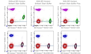
For optimal and reproducible results, BD Horizon Brilliant Stain Buffer should be used anytime two or more BD Horizon Brilliant dyes are used in the same experiment. Fluorescent dye interactions may cause staining artifacts which may affect data interpretation. The BD Horizon Brilliant Stain Buffer was designed to minimize these interactions. More information can be found in the Technical Data Sheet of the BD Horizon Brilliant Stain Buffer (Cat. No. 563794/566349) or the BD Horizon Brilliant Stain Buffer Plus (Cat. No. 566385).
Note: When using high concentrations of antibody, background binding of this dye to erythroid cell subsets (mature erythrocytes and precursors) has been observed. For researchers studying these cell populations, or in cases where light scatter gating does not adequately exclude these cells from the analysis, this background may be an important factor to consider when selecting reagents for panel(s).
Product Notices
- Since applications vary, each investigator should titrate the reagent to obtain optimal results.
- An isotype control should be used at the same concentration as the antibody of interest.
- Caution: Sodium azide yields highly toxic hydrazoic acid under acidic conditions. Dilute azide compounds in running water before discarding to avoid accumulation of potentially explosive deposits in plumbing.
- For fluorochrome spectra and suitable instrument settings, please refer to our Multicolor Flow Cytometry web page at www.bdbiosciences.com/colors.
- BD Horizon Brilliant Ultraviolet 737 is covered by one or more of the following US patents: 8,110,673; 8,158,444; 8,227,187; 8,575,303; 8,354,239.
- BD Horizon Brilliant Stain Buffer is covered by one or more of the following US patents: 8,110,673; 8,158,444; 8,575,303; 8,354,239.
- Please refer to http://regdocs.bd.com to access safety data sheets (SDS).
- Please refer to www.bdbiosciences.com/us/s/resources for technical protocols.
関連製品


The M1/69 monoclonal antibody specifically binds to CD24 (Heat-Stable Antigen, HSA or HsAg), a variably glycosylated, glycosyl-phosphatidylinositol-anchored membrane protein expressed on erythrocytes, granulocytes, monocytes, lymphocytes, and neurons. Hematopoietic stem cells of the embryonic yolk sac and fetal liver express CD24. Levels of expression of CD24 vary during differentiation of the T and B cell lineages. In the bone marrow, hematopoietic progenitors acquire CD24 expression upon commitment to the B-lymphocyte lineage. Immature B cells in the bone marrow express low CD24 levels whereas peripheral B lymphocytes express intermediate to high levels of CD24. The level of CD24 expression has been reported to rise upon activation of splenic B cells with LPS, but not with CD154 (CD40 Ligand). The majority of thymocytes express high levels of CD24, while most mature thymic and peripheral T lymphocytes do not express CD24. In contrast, TCR-bearing thymocytes which emigrate to the spleen are CD24+. Dendritic cells of the thymus, spleen, liver, and epidermal Langerhans cells have also been reported to express CD24. CD24 is not expressed by NK cells, as determined by staining with J11d mAb (Cat. No. 553146). CD24 is involved in the costimulation of CD4+ T cells by B cells, it is a co-inducer of in vitro thymocyte maturation, and it is a ligand of CD62P (P-selectin). While the monoclonal antibodies 30-F1, M1/69, and J11d all react with CD24, they show subtle differences in the level of staining of different lymphocyte populations. When possible, investigators should continue to use the same monoclonal anti-CD24 antibody as used in previous studies.
The antibody was conjugated to BD Horizon BUV737 which is part of the BD Horizon Brilliant™ Ultraviolet family of dyes. This dye is a tandem fluorochrome with an Ex Max near 350 nm and an Em Max near 737 nm. BD Horizon Brilliant BUV737 can be excited by the ultraviolet laser (355 nm) and detected with a 740/35 nm filter. Due to the excitation of the acceptor dye by the red laser line, there may be significant spillover into red laser detectors with filters in the 700-720 nm range.

Development References (11)
-
Allman DM, Ferguson SE, Cancro MP. Peripheral B cell maturation. I. Immature peripheral B cells in adults are heat-stable antigenhi and exhibit unique signaling characteristics. J Immunol. 1992; 149(8):2533-2540. (Clone-specific: Flow cytometry, Fluorescence activated cell sorting). View Reference
-
Allman DM, Ferguson SE, Lentz VM, Cancro MP. Peripheral B cell maturation. II. Heat-stable antigen(hi) splenic B cells are an immature developmental intermediate in the production of long-lived marrow-derived B cells. J Immunol. 1993; 151(9):4431-4444. (Clone-specific: Flow cytometry, Fluorescence activated cell sorting). View Reference
-
Alterman LA, Crispe IN, Kinnon C. Characterization of the murine heat-stable antigen: an hematolymphoid differentiation antigen defined by the J11d, M1/69 and B2A2 antibodies. Eur J Immunol. 1990; 20(7):1597-1602. (Clone-specific: Flow cytometry). View Reference
-
Cibotti R, Punt JA, Dash KS, Sharrow SO, Singer A. Surface molecules that drive T cell development in vitro in the absence of thymic epithelium and in the absence of lineage-specific signals. Immunity. 1997; 6(3):245-255. (Clone-specific: (Co)-stimulation, Flow cytometry). View Reference
-
Crispe IN, Bevan MJ. Expression and functional significance of the J11d marker on mouse thymocytes. J Immunol. 1987 April; 138(7):2013-2018. (Clone-specific: Flow cytometry). View Reference
-
Milstein C, Galfre G, Secher DS, Springer T. Monoclonal antibodies and cell surface antigens. Ciba Found Symp. 1979; 66:251-276. (Clone-specific: Flow cytometry). View Reference
-
Springer T, Galfre G, Secher D, Milstein C. Monoclonal xenogeneic antibodies to mouse leukocyte antigens: identification of macrophage-specific and other differentiation antigens. Curr Top Microbiol Immunol. 1978; 81:45-50. (Immunogen: Radioimmunoassay). View Reference
-
Springer T, Galfre G, Secher DS, Milstein C. Monoclonal xenogeneic antibodies to murine cell surface antigens: identification of novel leukocyte differentiation antigens. Eur J Immunol. 1978; 8(8):539-551. (Immunogen: Blocking, Radioimmunoassay). View Reference
-
Stall AM, Wells SM. FACS analysis of murine B-cell populations. In: Herzenberg LA, Weir DM, Blackwell C, ed. Weir's Handbook of Experimental Immunology. Blackwell Science Publishers; 1997:63.1-63.17.
-
Veillette A, Zuniga-Pflucker JC, Bolen JB, Kruisbeek AM. Engagement of CD4 and CD8 expressed on immature thymocytes induces activation of intracellular tyrosine phosphorylation pathways. J Exp Med. 1989; 170(5):1671-1680. (Clone-specific: Cell separation, Cytotoxicity). View Reference
-
Wenger RH, Rochelle JM, Seldin MF, Kohler G, Nielsen PJ. The heat stable antigen (mouse CD24) gene is differentially regulated but has a housekeeping promoter. J Biol Chem. 1993; 268(31):23345-23352. (Biology). View Reference
Please refer to Support Documents for Quality Certificates
Global - Refer to manufacturer's instructions for use and related User Manuals and Technical data sheets before using this products as described
Comparisons, where applicable, are made against older BD Technology, manual methods or are general performance claims. Comparisons are not made against non-BD technologies, unless otherwise noted.
For Research Use Only. Not for use in diagnostic or therapeutic procedures.
Report a Site Issue
This form is intended to help us improve our website experience. For other support, please visit our Contact Us page.