-
抗体試薬
- フローサイトメトリー用試薬
-
ウェスタンブロッティング抗体試薬
- イムノアッセイ試薬
-
シングルセル試薬
- BD® AbSeq Assay | シングルセル試薬
- BD Rhapsody™ Accessory Kits | シングルセル試薬
- BD® Single-Cell Multiplexing Kit | シングルセル試薬
- BD Rhapsody™ Targeted mRNA Kits | シングルセル試薬
- BD Rhapsody™ Whole Transcriptome Analysis (WTA) Amplification Kit | シングルセル試薬
- BD Rhapsody™ TCR/BCR Profiling Assays (VDJ Assays) | シングルセル試薬
- BD® OMICS-Guard Sample Preservation Buffer
-
細胞機能評価のための試薬
-
顕微鏡・イメージング用試薬
-
細胞調製・分離試薬
-
- BD® AbSeq Assay | シングルセル試薬
- BD Rhapsody™ Accessory Kits | シングルセル試薬
- BD® Single-Cell Multiplexing Kit | シングルセル試薬
- BD Rhapsody™ Targeted mRNA Kits | シングルセル試薬
- BD Rhapsody™ Whole Transcriptome Analysis (WTA) Amplification Kit | シングルセル試薬
- BD Rhapsody™ TCR/BCR Profiling Assays (VDJ Assays) | シングルセル試薬
- BD® OMICS-Guard Sample Preservation Buffer
- Japan (Japanese)
-
Change country/language
Old Browser
Looks like you're visiting us from {countryName}.
Would you like to stay on the current country site or be switched to your country?


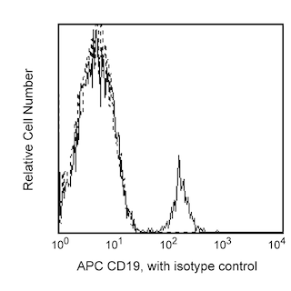
Two-color flow cytometric analysis of human CD307c expression on human peripheral blood lymphocytes. Whole blood was stained with APC Mouse Anti-Human CD19 antibody (Cat. No. 555415/561742), and either Biotin Mouse IgG2b, κ Isotype Control (Cat. No. 565415; Left Plot) or Biotin Mouse Anti-Human CD307c antibody (Cat. No. 565056; Right Plot). The cells were counterstained with PE Streptavidin (Cat. No. 554061). Erythrocytes were lysed with BD FACS Lysing Solution (Cat. No. 349202). Two-color flow cytometric contour plots showing the correlated expression of CD307c (or Ig Isotype control staining) versus CD19 were derived from gated events with the forward and side light-scatter characteristics of intact lymphocytes. Flow cytometric analysis was performed using a BD™ LSR II Flow Cytometer System.


BD Pharmingen™ Biotin Mouse Anti-Human CD307c

Regulatory Statusの凡例
Any use of products other than the permitted use without the express written authorization of Becton, Dickinson and Company is strictly prohibited.
Preparation and Storage
Product Notices
- This reagent has been pre-diluted for use at the recommended Volume per Test. We typically use 1 × 10^6 cells in a 100-µl experimental sample (a test).
- An isotype control should be used at the same concentration as the antibody of interest.
- Caution: Sodium azide yields highly toxic hydrazoic acid under acidic conditions. Dilute azide compounds in running water before discarding to avoid accumulation of potentially explosive deposits in plumbing.
- Source of all serum proteins is from USDA inspected abattoirs located in the United States.
- For fluorochrome spectra and suitable instrument settings, please refer to our Multicolor Flow Cytometry web page at www.bdbiosciences.com/colors.
- Please refer to www.bdbiosciences.com/us/s/resources for technical protocols.
関連製品


.png?imwidth=320)

The H5 monoclonal antibody specifically binds to CD307c which is also known as Fc receptor-like 3 (FCRL3), Immune receptor translocation-associated protein 3 (IRTA3), IFGP family protein 3 (IFGP3), and SH2 domain-containing phosphatase anchor protein 2 (SPAP2). The FCRL3 gene is present in humans but not in mice. CD307c is a type I transmembrane glycoprotein that belongs to the FCRL family within the Ig gene superfamily. CD307c shares sequence homology with classical the Fc receptors. CD307c is expressed on subsets of B cells, plasma cells, NK cells, CD8+ T cells, and CD4+ natural T regulatory cells. CD307c isoforms contain multiple extracellular Ig domains and immunoreceptor-tyrosine activation (ITAM) and inhibitory (ITIM) motifs in their intracellular domains. CD307c may be involved in the regulation of immune responses. Genetic variation in FCRL3 has been associated with susceptibility to certain autoimmune diseases.
Development References (6)
-
Kochi Y, Myouzen K, Yamada R, et al. FCRL3, an autoimmune susceptibility gene, has inhibitory potential on B-cell receptor-mediated signaling. J Immunol. 2009; 183(9):5502-5510. (Biology). View Reference
-
Llinas L, Lazaro A, de Salort J, Matesanz-Isabel J, Sintes J, Engel P. Expression profiles of novel cell surface molecules on B-cell subsets and plasma cells as analyzed by flow cytometry. Immunol Lett. 2011; 134(2):113-121. (Clone-specific: Flow cytometry). View Reference
-
Matesanz-Isabel J, Sintes J, Llinas L, de Salort J, Lazaro A, Engel P. New B-cell CD molecules. Immunol Lett. 2011; 134(2):104-112. (Clone-specific: Flow cytometry). View Reference
-
Nagata S, Ise T, Pastan I. Fc receptor-like 3 protein expressed on IL-2 nonresponsive subset of human regulatory T cells. J Immunol. 2009; 182(12):7518-7526. (Immunogen: ELISA, Flow cytometry, Fluorescence activated cell sorting, Functional assay). View Reference
-
Polson AG, Zheng B, Elkins K, et al. Expression pattern of the human FcRH/IRTA receptors in normal tissue and in B-chronic lymphocytic leukemia. Int Immunol. 2006; 18(9):1363-1373. (Biology). View Reference
-
Swainson LA, Mold JE, Bajpai UD, McCune JM. Expression of the autoimmune susceptibility gene FcRL3 on human regulatory T cells is associated with dysfunction and high levels of programmed cell death-1. J Immunol. 2010; 184(7):3639-3647. (Biology). View Reference
Please refer to Support Documents for Quality Certificates
Global - Refer to manufacturer's instructions for use and related User Manuals and Technical data sheets before using this products as described
Comparisons, where applicable, are made against older BD Technology, manual methods or are general performance claims. Comparisons are not made against non-BD technologies, unless otherwise noted.
For Research Use Only. Not for use in diagnostic or therapeutic procedures.
Report a Site Issue
This form is intended to help us improve our website experience. For other support, please visit our Contact Us page.