

Preparation and Storage
Product Notices
- Since applications vary, each investigator should titrate the reagent to obtain optimal results.
- An isotype control should be used at the same concentration as the antibody of interest.
- Caution: Sodium azide yields highly toxic hydrazoic acid under acidic conditions. Dilute azide compounds in running water before discarding to avoid accumulation of potentially explosive deposits in plumbing.
- The Alexa Fluor®, Pacific Blue™, and Cascade Blue® dye antibody conjugates in this product are sold under license from Molecular Probes, Inc. for research use only, excluding use in combination with microarrays, or as analyte specific reagents. The Alexa Fluor® dyes (except for Alexa Fluor® 430), Pacific Blue™ dye, and Cascade Blue® dye are covered by pending and issued patents.
- Alexa Fluor® is a registered trademark of Molecular Probes, Inc., Eugene, OR.
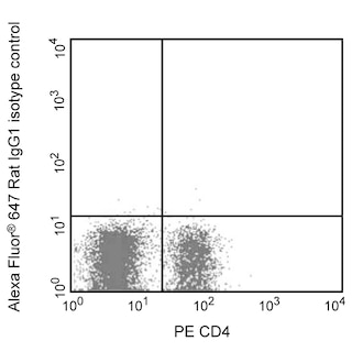
- Alexa Fluor® 647 fluorochrome emission is collected at the same instrument settings as for allophycocyanin (APC).
- For fluorochrome spectra and suitable instrument settings, please refer to our Multicolor Flow Cytometry web page at www.bdbiosciences.com/colors.
- Please refer to www.bdbiosciences.com/us/s/resources for technical protocols.
関連製品


The M3/84 monoclonal antibody specifically binds to CD107b which is also known as Mac-3, Lysosome-associated membrane protein 2 (LAMP-2/Lamp2/Lamp II), and Lysosomal membrane glycoprotein type B (LGP-B). CD107b is a single-pass type I transmembrane glycoprotein that constitutes a major integral membrane protein of lysosomes and may play a role in lysosomal function. CD107b is also expressed on the surface of mouse mononuclear phagocytes. Surface expression of the 92-110-kDa glycoprotein antigen increases during differentiation of monocytes to activated macrophages and may play a role in adhesion. The M3/84 mAb can detect CD107b antigen on tissue macrophages, thioglycollate-elicited peritoneal macrophages, and some myeloid cell lines, but not on lymphocytes or monocytes. In the bone marrow, very few cells display CD107b antigen on the surface, but a large proportion express cytoplasmic CD107b. The M3/84 antibody has also been reported to stain dendritic cells and endothelium in sections of thymus (both medulla and cortex), lymph nodes, spleen (white pulp), and gut-associated lymphoid tissue.
Development References (6)
-
Chen JW, Murphy TL, Willingham MC, Pastan I, August JT. Identification of two lysosomal membrane glycoproteins. J Cell Biol. 1985; 101(1):85-95. (Clone-specific: Immunoprecipitation). View Reference
-
Flotte TJ, Springer TA, Thorbecke GJ. Dendritic cell and macrophage staining by monoclonal antibodies in tissue sections and epidermal sheets. Am J Pathol. 1983; 111(1):112-124. (Clone-specific: Immunohistochemistry). View Reference
-
Ho MK, Springer TA. Tissue distribution, structural characterization, and biosynthesis of Mac-3, a macrophage surface glycoprotein exhibiting molecular weight heterogeneity. J Biol Chem. 1983; 285(1):636-642. (Clone-specific: Immunoprecipitation). View Reference
-
Springer TA. Cell-surface differentiation in the mouse. Characterization of "jumping" and "lineage" antigens using xenogeneic rat monoclonal antibodies. In: Kennett RH, McKearn TJ, Bechtol KB, ed. Monoclonal antibodies. Hybridomas: A new dimension in biological analyses. New York and London: Plenum Press; 1980:185-217.
-
Springer TA. Monoclonal antibody analysis of complex biological systems. Combination of cell hybridization and immunoadsorbents in a novel cascade procedure and its application to the macrophage cell surface. J Biol Chem. 1981; 256(8):3833-3839. (Immunogen: Immunoprecipitation). View Reference
-
Walker EB, Akporiaye ET, Warner NL, Stewart CC. Characterization of subsets of bone marrow-derived macrophages by flow cytometry analysis. J Leukoc Biol. 1985; 37(2):121-136. (Biology). View Reference
Please refer to Support Documents for Quality Certificates
Global - Refer to manufacturer's instructions for use and related User Manuals and Technical data sheets before using this products as described
Comparisons, where applicable, are made against older BD Technology, manual methods or are general performance claims. Comparisons are not made against non-BD technologies, unless otherwise noted.
For Research Use Only. Not for use in diagnostic or therapeutic procedures.